| Posted: Sep 11, 2015 |
Modeling self-organization in biological pattern formation(Nanowerk News) While it has been known for many years that biological pattern formation relies on the self-organization of molecular components into specific patterns, the mechanisms involved have proved difficult to elucidate. Now, researchers at RIKEN and Institut Curie in France have developed and experimentally verified a mathematical model that describes self-organization of actin filaments in developing tracheal tubules of flies, illuminating the mechanisms involved ("Cortical instability drives periodic supracellular actin pattern formation in epithelial tubes"). |
| The team investigated the respiratory tracheal system of the fruit fly Drosophila and focused on two proteins—actin, that polymerizes into filaments that form the skeleton of cells, and myosin, a ‘molecular motor’ that binds to actin filaments and moves them. In Drosophila embryos, these proteins initially form a uniform gel on the inner surface of the outer layer of tracheal tubules (the cortex), but during development they self-organize into ring-like patterns (Fig.). |
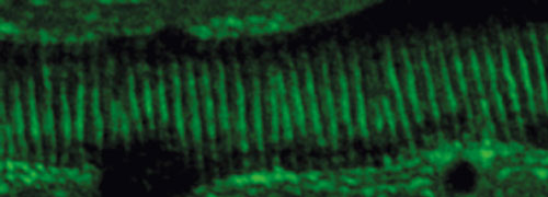 |
| Equally spaced actin rings form in the developing tracheal tubules of fruit flies. |
| “Actin patterns often prefigure important cell biological activities, such as cell movement and cell adhesion,” explains Shigeo Hayashi, who led the study and is from the RIKEN Center for Developmental Biology. “In Drosophila, actin rings form the prepattern for forming the exoskeleton.” |
| Based on the observation that the formation of actin rings coincided with circumferential assembly of actin filaments, the researchers constructed a mathematical model of the interactions leading to actin ring formation. They performed experimental measurements to determine the values of the model’s parameters and then used the model to make testable predictions about the self-organization process. |
| One of the model’s predictions was that pattern formation depends on the contractility of myosin. To test this, the researchers incubated Drosophila embryos with a drug that reduces the contractility of myosin. As predicted by the model, the drug prevented actin rings from forming. |
| Another prediction was that the properties of the actin–myosin gel determine the spacing of the actin rings. The team genetically modified Drosophila embryos to weaken the interactions between the actin–myosin gel and the embryo cortex. This reduction in molecular friction led to wider spacing of the actin rings. |
| The consensus between experimental results and the model’s predictions confirms that the model accurately describes the process and that the self-organization of actin rings depends on the availability of actin and myosin, the contractility of myosin, and the strength of molecular interactions. |
| “Self-organization is the core principle of biological pattern emergence,” says Hayashi, “and our model explains not only intracellular actin pattern formation, but also tissue-level coordination of actin pattern formation in response to extracellular clues.” |
| Source: RIKEN |

