| Mar 21, 2024 |
New method for analysing nanoporous materials
(Nanowerk News) In addition to their main components, the properties of crystalline and nanoporous materials often depend crucially on guest atoms or ions that are embedded in the tiny pores of their lattice structure. This applies to high-tech materials used in sensor or separation technology as well as to natural materials.
|
|
The bluish gemstone aquamarine, for example, would be colourless without such guest components. Determining the type and position of guest components is difficult, as many materials react sensitively to the radiation emissions from electron microscopes.
|
|
Thanks to a new method developed by a team led by Daniel Knez and Ferdinand Hofer at the Institute of Electron Microscopy and Nanoanalysis at Graz University of Technology (TU Graz), this can now be done with less radiation and is therefore much easier.
|
|
“The uniqueness of our method lies in the fact that we can determine the three-dimensional distribution of ions in crystal channels or nanopores based on a single electron microscope image,” says Daniel Knez.
|
|
The findings have been published in Communications Materials ("Three-dimensional distribution of individual atoms in the channels of beryl").
|
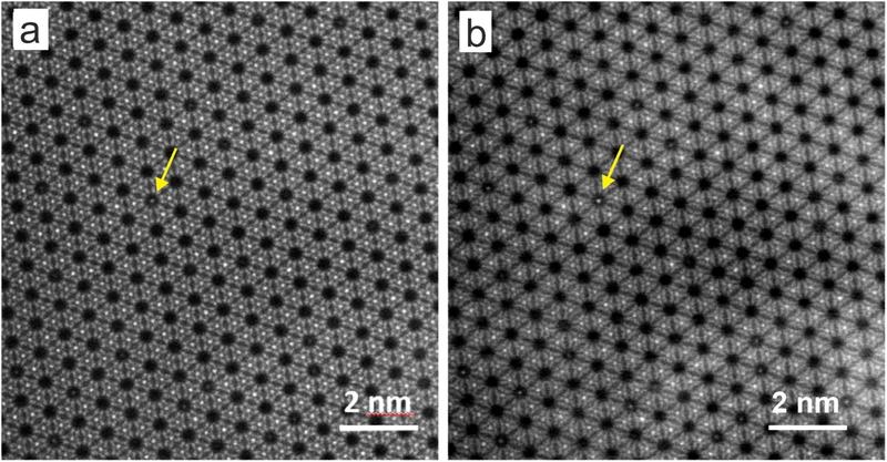 |
| An electron microscope image of an aquamarine. The yellow arrows mark caesium ions in the crystal pores. (Image: FELMI-ZFE)
|
The mysterious blue colour of aquamarine
|
|
The researchers developed their method while analysing the gemstone aquamarine. Until now, it was not known where exactly the iron that lends the stone its blue colour is positioned in the crystal. One hypothesis was that individual iron atoms are stuck in the pores and create this effect from there. But this has now been refuted.
|
|
In their experiments, the researchers have established beyond doubt that there is no iron in the pores, but instead caesium ions. The colour-conferring iron atoms are located in close proximity to the caesium ions, but are integrated into the columns of the crystal lattice.
|
A single image with atomic resolution as a basis
|
|
For their experiments, the researchers recorded a so-called Z-contrast image of the aquamarine crystal at atomic resolution using the ASTEM microscope, a scanning transmission electron microscope.
|
|
The electron beam of the ASTEM microscope is focused on the surface of the crystal sample, also penetrating into the pores of the material. If it hits ions stored there, they appear as bright dots in the image. Based on the strength of the contrast with empty pores and the neighbouring lattice structures, the researchers can determine the type of embedded ions and also estimate how deep they are located in the pores. These data were statistically analysed and compared with a large number of simulations of the crystal structure in order to be able to estimate the various factors influencing the measured signal.
|
Innovative method opens up new possibilities for materials science
|
|
In addition to basic research, the new method is also suitable for the targeted development of new materials. “Our method can be used to precisely determine the position of doping elements, i.e. targeted function-controlling additives, in nanoporous materials such as zeolites or metal-organic framework compounds,” says Ferdinand Hofer. This facilitates the optimisation of (single-atom) catalysts and solid-state electrolytes in future batteries or the development of biomedical applications for controlling drug uptake.
|

