| Aug 21, 2025 |
Angstrom microscopy reveals hidden states of single membrane proteinsCryogenic optical microscopy uncovers multiple conformations of PIEZO1 in cell membranes, offering atomic-scale insights into how cells sense force.(Nanowerk News) Our ability to sense touch and pressure relies on membrane proteins that convert mechanical forces into biological signals. A new study in Science Advances ("Cryo–light microscopy with angstrom precision deciphers structural conformations of PIEZO1 in its native state") from the Max Planck Institute for the Science of Light (MPL) demonstrates that cryogenic optical microscopy can capture such proteins in unprecedented detail, reaching ångström precision within their native cellular membranes. |
| The team focused on PIEZO1, a mechanosensitive ion channel that bends the cell membrane into a dome-like shape. Using fluorescent labeling and rapid shock-freezing to preserve the protein’s natural environment, the researchers built a cryogenic microscope capable of pinpointing single fluorescent markers with atomic-scale accuracy. |
| This approach revealed three distinct structural states of PIEZO1, ranging from highly curved to nearly flat conformations. The results provide quantitative evidence for how the protein’s blades flex and rotate, a motion thought to underlie its gating mechanism that opens the channel to ions. Importantly, the study shows that PIEZO1 in its native membrane can assume configurations not fully captured by earlier cryo-electron microscopy. |
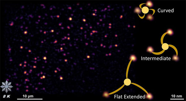 |
| Fluorescent image of PIEZO1 in native cell membrane (left) and an artistic representation of its conformational states resolved with Ångström precision (right). (Image: Hisham Mazal) |
| The method also allowed the team to probe a mutant form of PIEZO1 that favors an open state, confirming structural shifts predicted by electrophysiological experiments. By mapping these conformations at the single-molecule level, the researchers demonstrate that cryogenic optical microscopy can resolve structural heterogeneity without the averaging required in traditional electron microscopy. |
| “This development opens a new frontier in structural biology,” says Prof. Vahid Sandoghdar, director at MPL. “It provides direct access to the conformational states of proteins inside membranes, helping us understand the molecular mechanics of life at atomic precision.” |
| The researchers envision combining this optical technique with cryo-EM to achieve even deeper insights. Their results mark a step toward a quantitative picture of how proteins respond to mechanical forces—knowledge that could illuminate not only touch and pain sensation, but also mechanobiology across many systems. |
| Source: Max Planck Institute for the Science of Light (Note: Content may be edited for style and length) |

