| Posted: June 23, 2009 |
New electron microscopy images reveal the assembly of HIV |
| (Nanowerk News) Scientists at the European Molecular Biology Laboratory (EMBL) and the University Clinic Heidelberg, Germany, have produced a three-dimensional reconstruction of HIV (Human Immunodeficiency Virus), which shows the structure of the immature form of the virus at unprecedented detail. Immature HIV is a precursor of the infectious virus, which can cause AIDS. The study, published in the 22-26 June online edition of PNAS ("Structure and assembly of immature HIV"), describes how the protein coat that packages the virus' genetic material assembles in human cells. Drugs that block this assembly process and prevent the virus from maturing into its infectious form are considered a promising therapeutic approach. |
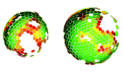 |
| Lattice maps for immature HIV particles. The 3D computer reconstruction shows the immature Gag lattice of HIV that matures to form the protein shell of the infecious virus. Maps are shown in perspective such that hexamers on the rear surface of the particle appear smaller. The side of the particle toward the viewer lacks ordered Gag. (Image: John Briggs/EMBL) |
| HIV consists of an RNA molecule that carries the genetic information of the virus and is surrounded by protective protein and membrane layers. During infection the virus deposits its genetic material into a human cell where it reprogrammes the host cell machinery to generate many copies of the viral genome and initiates the production of a viral protein called Gag. In the immature virus, many copies of Gag interact to form a roughly spherical lattice that encloses the virus' genetic material. The virus then leaves the cell with the help of proteins of the host and infects new cells. |
| Using a method called cryoelectron tomography researchers in the groups of John Briggs at EMBL and Hans-Georg Kräusslich at the University Clinic Heidelberg generated the as yet highest resolution 3D computer reconstruction images of the immature Gag lattice. The results suggest a simple model of HIV formation in human cells: multiple Gag proteins interact to form a hexameric lattice that grows with an inherent curvature and that incorporates new proteins stochastically. Several further steps in which Gag is cleaved by an enzyme are necessary to transform this immature lattice into its mature, infectious form. |
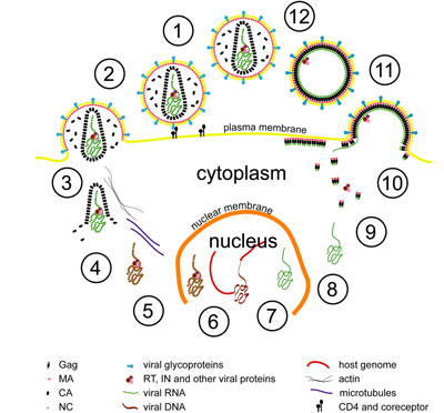 |
| Simplified representation of HIV’s lifecycle: The HIV lifecycle begins with the interaction of a virus particle with a receptor on the surface of a cell (step 1), which leads to fusion of the viral and cellular membranes (step 2) and deposition of the viral contents into the cell (step 3). The viral RNA genome is reverse transcribed resulting in DNA copy, which is imported into the nucleus (step 5). Within the nucleus, the viral DNA is integrated into the host cell genome (step 6). The virus may now enter a period of latency. The late stages of the viral lifecycle begin when the viral genome is transcribed from within the host genome (step 7), exported from the nucleus (step 8) and translation of the viral proteins by host cell machinery begins (step 9). The major structural protein, Gag, is transported to the plasma membrane where it directs assembly of the viral coat, and incorporates other viral proteins and the viral genome (step 10). The virus buds through the cell membrane. |
| Briggs and his team are now working on producing an even higher resolution structure of the protein lattice to gain a more detailed understanding of the virus' assembly and maturation processes, which may eventually help to find weak points that could be targeted by drugs. |
| Cryoelectron tomography is a technique with which a sample is instantly frozen in its natural state and then examined with an electron microscope. Images are taken from different directions and assembled into an accurate 3D reconstruction by a computer. |
| Source: European Molecular Biology Laboratory (EMBL) |

