| Posted: June 30, 2009 |
The sound of light: Innovative technology shatters the barriers of modern light microscopy |
|
(Nanowerk News) Since the dawn of the microscope scientists have been using light to scrutinize thin sections of tissue to ascertain whether they are healthy or diseased or to investigate cell function. However, the penetration limits for this kind of examination lie between half a millimeter and one millimeter of tissue. In thicker layers light is diffused so strongly that all useful details are obscured.
|
|
Together with his research team, Professor Vasilis Ntziachristos, director of the Institute of Biological and Medical Imaging of the Helmholtz Zentrum München – German Research Center for Environmental Health and chair for biological imaging at the Technische Universität München, has now broken through this barrier and rendered three-dimensional images through at least six millimeters of tissue, allowing whole-body visualization of adult zebra fish (Multispectral opto-acoustic tomography of deep-seated fluorescent proteins in vivo).
|
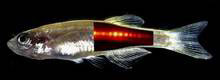 |
| Light and ultrasound can be used to visualize the red fluorescent spinal column of a live fish: Multi-spectral opto-acoustic tomography or MSOT allows the investigation of subcellular processes in live organisms (Helmholtz Zentrum München / TU München, montage)
|
|
To achieve this feat, Prof. Ntziachristos and his team made light audible. They illuminated the fish from multiple angles using flashes of laser light that are absorbed by fluorescent pigments in the tissue of the genetically modified fish. The fluorescent pigments absorb the light, a process that causes slight local increases temperature, which in turn result in tiny local volume expansions. This happens very quickly and creates small shock waves. In effect, the short laser pulse gives rise to an ultrasound wave that the researchers pick up with an ultrasound microphone.
|
|
The real power of the technique, however, lies in specially developed mathematical formulas used to analyze the resulting acoustic patterns. An attached computer uses these formulas to evaluate and interpret the specific distortions caused by scales, muscles, bones and internal organs to generate a three-dimensional image.
|
|
The result of this “multi-spectral opto-acoustic tomography”, or MSOT, is an image with a striking spatial resolution better than 40 micrometers (four hundredths of a millimeter). And best of all, the sedated fish wakes up and recovers without harm following the procedure.
|
|
Dr. Daniel Razansky, who played a pivotal role in developing the method, says, "This opens the door to a whole new universe of research. For the first time, biologists will be able to optically follow the development of organs, cellular function and genetic expression through several millimeters to centimeters of tissue.”
|
|
|
|
In the past, understanding the evolution of development or of disease required numerous animals to be sacrificed. With a plethora of fluorochrome pigments to choose from – including pigments using the fluorescence protein technology for which a Nobel Prize was awarded in 2008 and clinically approved fluorescent agents – observing metabolic and molecular processes in all kinds of living organisms, from fish to mice and humans, will be possible. The fruits of pharmaceutical research can also be harvested faster since the molecular effects of new treatments can be observed in the same animals over an extended period of time.
|
|
Bio-engineer Ntziachristos is convinced that, “MSOT can truly revolutionize biomedical research, drug discovery and healthcare. Since MSOT allows optical and fluorescence imaging of tissue to a depth of several centimeters, it could become the method of choice for imaging cellular and subcellular processes throughout entire living tissues.”
|

