| Posted: November 30, 2009 |
New research offers clues to how shells grow in nature |
|
(Nanowerk News) Single crystals of the mineral calcite -- the chief material in limestone -- are predictable, homogeneous and, well, a little boring.
|
|
But scientists have long marveled at how biological crystals of calcite grow together with other organic materials to form, for example, shells and sea urchin spines. Biologists and materials scientists would love to know exactly how to re-create such natural composites in the lab.
|
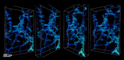 |
| This image shows the 3-D nanoscale structure of the inside of a synthetic calcite crystal, which was grown in an agarose gel. In this environment, the crystal grows around the polymeric fibers. The structure gives insight into the formation of single crystals by biological organisms.
|
|
Lara Estroff, assistant professor of materials science and engineering, and colleagues have taken a deep, detailed look at the way lab-created calcite crystals, similar to those found in nature, grow in tandem with proteins and other large molecules, and they report their findings in the Nov. 27 issue of the journal Science. The journal also features the work as a "Perspective" written by Kansas State University's Mark Hollingsworth.
|
|
"We knew the organics were in there, but what no one had been able to do up until now was actually see what that organic-inorganic interface looked like," said Estroff, whose lab focuses on the synthesis and characterization of bio-inspired materials.
|
|
Estroff and graduate student Hanying Li grew samples of calcite in a hydrogel called agarose -- a common dessert additive -- that mimics the way calcite grows in living things. In previously published work, Li and Estroff had determined that this gel environment made the crystals grow very differently than in solution.
|
|
In collaboration with associate professor of applied and engineering physics David Muller and physics graduate student Huolin Xin, the researchers prepared their crystals with Focused Ion Beam (FIB) technology, which uses high-energy ions to slice samples thin enough for an electron beam to pass through for imaging.
|
|
Scanning transmission electron microscopy methods developed by Xin and Muller revealed detailed, three-dimensional pictures of the internal structure of calcite crystals grown in the gel. They found that the crystals trap large molecules by growing around them.
|
|
Studying this complex natural process may be a key step toward giving materials scientists like Estroff clues on how to make and manipulate nature-inspired composite materials. Applications could range from electronics to photovoltaics to completely new classes of materials.
|

