| Mar 11, 2011 |
Turn your smartphone into a biomedical device
|
|
(Nanowerk News) In a new, free-access paper in PloS one ("Cell-Phone-Based Platform for Biomedical Device Development and Education Applications"), researchers propose to take advantage of the rapid improvements in commercial CMOS sensors and microscopic optics driven by the cell-phone industry to develop two common biomedical devices, namely a microscope and spectrometer, that are available as simple and inexpensive add-ons to a commercial cell phone camera like Apple's iPhone.
|
|
While other researchers have demonstrated previously similar devices, in this report the attachments to the phone are much smaller, simpler, and very low cost while still maintaining an acceptable level of performance. Here, the team demonstrate their relevance in laboratory measurements as well as discuss their applications within the field of science education.
|
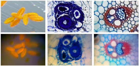 |
| Images of several commercially prepared microscope slides featuring stained samples. Top row, commercial microscope. Bottom row, cell phone microscope. Left column, pollen grains. Right two columns, plant stems.
|
|
Background
|
|
With health care costs increasing throughout the world, there is a pressing need for reducing the cost and complexity of biomedical devices. Additionally, with growing demand for high-quality health care in regions of the world where medical infrastructure is below levels found in developed countries, portable devices that can transmit relevant data to remote experts are likely to have a large impact on quantity and quality of care. To this end, several groups have focused on the development of low-cost and rapidly deployable technologies that address common diseases afflicting the third world and common tests performed in both hospital and field environments.
|
|
Cell phone cameras are certainly the most ubiquitous optical sensor in both the developed and developing worlds, and are attractive candidates for conversion to medical devices. Some work has already been directed towards this end, with several recent papers discussing the use of cell phones as diagnostic devices.
|
|
Researchers at UCLA have constructed a modified lensless cell phone that enables holography-based digital microscopy, while researchers at UC Berkeley have constructed a complex objective attachment that also transforms a cell phone into a microscope. Additionally, a patent was recently awarded for the use of a cell phone as a spectrometer. However, there is still a need for more research directed towards utilizing cell-phone cameras to record images or spectra of biological samples.
|
|
The use of low-quality, low-cost components makes sense in the context of visual pathologic inspection. In this application, trained professionals manually examine samples to observe tissue- and cellular-level disorders, often with the aid of optical dyes. In fact, the fundamental basis of pathologic diagnosis has remained essentially unchanged for more than 100 years, following the standardization of staining procedures such as hematoxylin and eosin for tissue sections and Wright-Giemsa staining for blood samples.
|
|
Additionally, the ability to cheaply and rapidly record diffuse reflectance spectra or fluorescence spectra also has the possibility to help with medical diagnosis. One example application is in the use of a spectrometer as a pulse oximeter, where the transmitted intensity through a finger is monitored and correlated through known absorption spectra to the concentration of oxy- and deoxy-hemoglobin. Additionally, a portable spectrometer might be used for the noninvasive detection of tumors, where it has been shown that tumors differ from surrounding healthy tissue by their increased autofluorescence and differing diffuse optical properties.
|

