| Jan 31, 2012 |
JPK reports on the current research activities of Dr Clemens Franz and his team at KIT |
| (Nanowerk News) Dr Clemens Franz leads a group of researchers at the DFG-Center for Functional Nanostructures at Karlsruhe Institute of Technology where he works on expanding the use of AFM for cell biological applications. AFM has a strong advantage over other microscopy techniques. Samples can be imaged directly without prior preparation, such as staining or fixation. This makes the technique extremely suitable for imaging biological samples as biological molecules or even living cells can be maintained under physiological conditions. The group also routinely uses AFM to characterize cell adhesion substrates. |
| Given that AFM cantilevers are ultrasoft springs, they can be used to measure inter- and even intramolecular bonds. To study cell adhesion, Dr Franz and his team often employ AFM-based single-cell force spectroscopy (SCFS). Here, a living cell is attached to the AFM cantilever and brought into contact with a substrate under defined contact conditions (contact force and time). With a force sensitivity spanning over four orders of magnitude, SCFS provides a unique opportunity to measure cellular adhesion forces from the single-molecule level to overall adhesion in the same experimental setup. AFM-based SCFS has provided important insight into molecular mechanisms involved in adhesion force generation, such as the transition from single-receptor mediated to cooperative receptor binding during the initial phase of cellular contact with an extracellular substrate. Furthermore, the adhesive properties of different surfaces can be accurately characterized. |
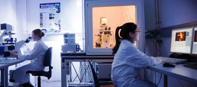 |
| Ramona Ring and Lu Dao of the Franz Group at Karlsruhe using JPK NanoWizard systems. |
| Their latest challenge is to combine AFM with advanced light microscopy techniques, such as laser scanning confocal or total internal reflection (TIRF) microscopy. In this way, the clustering of receptors can be followed in living cells by optical time-lapse-microscopy and directly correlated to the adhesion force information. The group now has built a functional TIRF/AFM setup. |
| The motivation for this work is to better understand fundamental mechanisms of cell adhesion, particularly initial adhesion events when cells first encounter components of the extracellular matrix (ECM). Through the use of micro- or nano-patterned artificial cell adhesion substrates, the goal is to emulate the natural cell environment and to manipulate cell adhesion. |
| Dr Franz describes why he likes to use the JPK AFM systems: "JPK's products are particularly suitable for biological research because of their relative ease of use; their good hardware/software integration; the availability of heated sample chambers and the possibility to image in fluid to keep biological sample functional. Furthermore, the JPK offers excellent integration of AFM and optical microscopy techniques (such as phase contrast, confocal or TIRF microscopy) while the unique 100 µm z-piezo pulling range (CellHesion® modules) are essential for complete cell-substrate separation in single-cell force spectroscopy." |
| For more details about JPK's specialist products and applications for the bio and nano sciences, please contact JPK on +49 30533112070, visit the web site: http://www.jpk.com/ or see more on Facebook: www.jpk.com/facebook. |
| Source: JPK (press release) |

