| Jan 31, 2012 |
Nanomedicine: "Russian doll" polymer vesicles mimic cell structure
|
|
(Nanowerk News) Nanomedicine faces two main challenges: controlling the synthesis of extremely small vectors containing one or several active ingredients and releasing these agents in the right place at the right time, in controlled forms and doses. Researchers from the Organic Polymer Chemistry Laboratory (CNRS/ Bordeaux 1 University/ Institut Polytechnique de Bordeaux) have recently encapsulated nanovesicles within slightly larger vesicles. This "Russian doll" structure mimics the organization of cell compartments. Reproducing it is a first major step towards triggering controlled reactions within the structure of the cell. This work is already opening up new possibilities in terms of multiple encapsulation, compartmentalized reactors and the administration of vectors via new delivery routes (e.g. oral absorption). These results are published on 27 January 2012, in Angewandte Chemie International Edition ("Polymersomes in Polymersomes: Multiple Loading and Permeability Control").
|
|
The principal nanovectors for drug delivery to have been studied so far are lipid vesicles or "liposomes". Analogs of these vectors based on polymers and known as "polymersomes" were discovered about 10 years ago. They have several advantages over liposomes: they are more stable and impermeable, they are more easily "functionalized" and "modulated" (it is possible, for example, to synthesize heat-sensitive polymers or polymers that recognize particular types of cells, such as tumor cells in particular). Over the last 10 years, the team coordinated by Sébastien Lecommandoux has been developing "intelligent" polymersomes from polypeptides whose properties and structures are analogous to those of viruses.
|
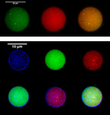 |
| Top, encapsulation of two types of internal polymersome populations, one in green and the other in red. Bottom, encapsulation in all the possible compartments: external membrane (blue), cavity of the external polymersome (green), internal polymersomes (red).
|
|
The researchers are now taking this biological mimicry and inspiration further, by encapsulating polymersomes within each other. This compartmentalization mimics the structure of cells, which are themselves composed of compartments (small internal organelles1, where thousands of interactions and reactions take place everyday) and a viscoelastic cytoplasm, providing the cell with a degree of mechanical stability. However, forming such encapsulated polymersomes in a controlled manner is no mean feat.
|
|
The scientists managed to do so through the use of a novel emulsion/centrifugation method that was quick, easy, required few reagents and proved highly effective. The team then used imaging with fluorescent markers to demonstrate the formation of structures in which polymersomes were encapsulated within each other. Controlling this compartmentalization makes it possible to envisage the encapsulation of several compounds (inside multiple internal polymersomes) within a single vector. This is what the researchers then went on to demonstrate: they encapsulated two different populations of internal polymersomes within a single larger polymersome. Their findings indicate that it should be possible to incorporate a much larger number of different vesicles within the vector. This is very promising for combined vectorization, in oncology for example, where the possibility of delivering different active ingredients (some of which may otherwise be incompatible) via a single vector would be a major advantage.
|
|
These novel structures could also be used as compartmentalized reactors, in catalysis or for biomedical applications. The researchers encapsulated three different fluorescent molecules2 (used as "model active ingredient molecules") in the three compartments comprised in these structures: the external polymersome membrane, the aqueous cavity of the external polymersome and the internal polymersome membrane3. Thus, it is now plausible to encapsulate different reagents in the various compartments of the polymersomes or to trigger different cascade reactions at will in these polymersomes.
|
|
In addition to providing improved protection for the encapsulated active ingredients, this packaging approach also facilitates control and allows a more precise modulation of the permeability properties of the vesicles. The researchers modeled this in an experiment involving the in-vitro release of an anticancer agent, doxorubicin (DOX), incorporated in internal encapsulated polymersomes. DOX was released about twice as rapidly from classical nanopolymersomes than from such polymersomes encapsulated within a larger external polymersome.
|
|
The researchers are the first to have achieved this type of multiple, controlled encapsulation in compartmentalized vesicles, especially polymers, that also mimic the cytoskeleton, thus reproducing the structure of the cell in its entirety4. The next step will be to use this system to trigger controlled chemical reactions in attoliter volumes (10-18 liters), in a confined environment. |
|
Notes
|
|
1- Small, differentiated internal structures (enclosed by a membrane) with specialized functions in living cells.
|
|
2- Three colors were used here: blue, green and red.
|
|
3- The internal polymersomes also have a cavity, but they are to small to allow the membrane to be distinguished from the cavity.
|
|
4- Polymersomes in "gelly" Polymersomes: towards structural cell mimicry. Maïté Marguet, Olivier Sandre, Sébastien Lecommandoux. Langmuir 2012 (DOI : la-2011-04018w).
|

