| May 22, 2012 |
Greater than the sum of their parts - functional molecular complexes
|
|
(Nanowerk News) For the first time, individual biomolecules have been assembled to form a molecular complex with its own unique function.
|
|
A few years ago, the journal Science published an article on a remarkable new method. Scientists had managed – as if using a construction crane – to pick up individual biomolecules and, with nanometer precision, to move them to a specific new location (see our Nanowerk Spotlight in this work: "Nanotechnology cut and paste with single molecules"). This technique, known as "single-molecule cut & paste," was developed by a working group led by NIM researcher Hermann Gaub of LMU Munich.
|
|
In their latest paper, Gaub and his colleagues report on how they used this method to construct a complex with its own unique function from two biomolecules. The biophysicists arranged two RNA segments to form a binding pocket that is specifically capable of binding malachite green dye. When the molecule's function is activated, the malachite green illuminates a thousand times more intensively than before, thereby signaling that all the molecules have found their proper place at the nano-construction site.
|
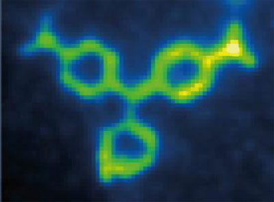 |
| Bottom up assembly of functional molecular ensembles with novel properties emerging from composition and arrangement of its constituents is a prime goal of nanotechnology. By single-molecule cut-and-paste we assembled binding sites for malachite green in a molecule-by-molecule assembly process from the two halves of a split aptamer. (© American Chemical Society)
|
|
The precise manipulation of individual atoms and molecules first began around 20 years ago. In a widely publicized feat of nano-engineering, scientists succeeded in arranging 35 xenon atoms so precisely that one could distinctly read the lettering "IBM" using a microscope. However, such experiments had to be conducted under extreme conditions – in an ultra-high vacuum and at a temperature of 4°K (approx. -270°C). It was impossible to use this method to arrange biomolecules, since their structure would be immediately destroyed.
|
|
In 2008, however, Hermann Gaub's team developed a technique that made it possible to pick up individual biomolecules from a surface and move them to another location. This method, known as "single-molecule cut & paste," functions under conditions compatible with life, i.e. at room temperature and ambient pressure. A scanning force microscope serves as the "crane," which grabs individual molecules with its nanometer-sized delicate tip.
|
|
In their new paper in the journal Nano Letters ("Functional Assembly of Aptamer Binding Sites by Single-Molecule Cut-and-Paste"), Gaub and his team show for the first time that it is possible to use this method to assemble a functional system from initially non-functional nano-building blocks. In this way, they furnish a compelling example of the dictum, "The whole is greater than the sum of its parts." As building blocks, Gaub and his team used RNA molecules that were merged into an "aptamer," i.e. a structure that is capable of recognizing and binding a target substance.
|
|
To assemble the molecules, the scientists used the fine tip of a scanning force microscope to pick up one RNA segment and transport it to a second RNA segment. Once a contact was formed between the two RNA strands, the aptamer then became functional: it formed a pocket structure into which the malachite green molecule fits exactly.
|
|
"It is important to have precise mechanical control over the assembly process. Only if the construction of the aptamer is successful do we see the malachite green molecule illuminate under the fluorescence microscope," said Mathias Strackharn, lead author of the article. "Thanks to this technique, we are now able to assemble systems whose natural function depends upon their configuration. Using the tools at our disposal, we can illuminate precisely how the molecules interact."
|

