| Posted: September 11, 2008 |
Researchers take 15 nanosecond snapshots of nanoscale transformations |
|
(Nanowerk News) Researchers have achieved a milestone in materials science and electron microscopy by taking a high-resolution snapshot of the transformation of nanoscale structures.
|
|
Using the Lab's Dynamic Transmission Electron Microscope (DTEM), Judy Kim and colleagues peered into the microstructure and properties of reactive multilayer foils (also known as nanolaminates) with 15-nanosecond-scale resolution.
|
|
"This is the first time that a detailed study of these reactive nanolaminates has exposed what is happening in the self-propagating high-temperature synthesis zone," Kim said.
|
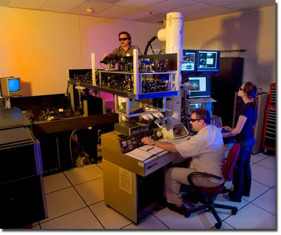 |
| From left: Curtis Brown, Thomas LaGrange and Judy Kim make adjustments to the dynamic transmission electron microscope. (Photo by Marcia Johnson/LLNL)
|
|
Time-resolved images of nanolaminates show a brief change in structure with a short cellular phase separation during cooling.
|
|
Observing short-lived behavior - how a chemical reaction, structural deformation or phase transformation occurs - is not easy, but is key to understanding many of the basic phenomena at the heart of chemistry, biology and materials science. The ability to directly observe and characterize these complex events leads to a fundamental understanding of properties such as reactivity, stability and strength, and helps in the design of new and improved materials and devices.
|
|
Transmission electron microscopy has evolved dramatically in recent years and can spatially resolve microstructural details of phase and structure, but it can't collect at times less than a millisecond.
|
|
That's where Livermore's DTEM comes in. It provides scientists with the ability to image transient behavior with an unprecedented combination of spatial and temporal resolution: nanometers and nanoseconds.
|
|
"Direct real-space observations of phase transformations on the nanosecond scale have allowed us to relate the formation mechanism in reactive multilayer foils to binary alloy solidification," Kim said. "This conclusion is based upon transient features that could not have been found using any other technique."
|
|
Because the team needed access to time and real-space that is impossible to study using traditional methods such as conventional in situ TEM (limited to video rates) or ultrafast diffraction (which presently lacks direct real-space imaging capabilities), the DTEM fit the bill.
|
|
"The ability to determine not only crystal structure, but also morphological evolution of dynamic events on the nanoscale has far-reaching implications for the study of materials science, non-equilibrium processes and the behavior of matter on very fine scales of length and time," Kim said.
|
|
Multilayer foils are layers of reactant materials that undergo exothermic, self-propagating reactions when layer mixing is caused by an external energy source. The foils show mobile, high-temperature reaction zones where atoms of adjoining layers diffuse across the interfaces. They are used as customized heat sources for rapid fuses, biological neutralization and joining materials via localized heating rather than global device heating.
|
|
The research appears in the Sept. 12 edition of the journal Science.
|

