| Feb 13, 2018 |
Chemists develop motion capture-like technology for tracking protein shape
|
|
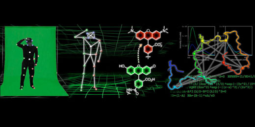
(Nanowerk News) In many modern animated movies, the trick to achieving realistic movements for individual characters and objects lies in motion-capture technology. This process often involves someone wearing a tracking suit covered in small, colored balls while a camera captures the position of those colored balls, which is then used to represent how the person is moving.
|
 |
|
Researchers at the University of Pennsylvania are developing a similar technology to obtain atomic-resolution "movies" that track how proteins fold and change shape. To generate these movies, the scientists label the protein with probes at many positions and observe the movement of those labels. The fluorescence data on the relative positions of the probes can then be used to construct computational models of the protein structure in atomic detail. This research could lead to improvements in drugs used to treat neurodegenerative diseases, as well as new methods of imaging that could lead to their earlier detection.
|
|
The research was multi-disciplinary effort led by E. James Petersson, associate professor of chemistry in Penn's School of Arts and Sciences, and graduate student Jack Ferrie. Elizabeth Rhoades and Zahra Fakhraii, both associate professors of chemistry at Penn, as well as Abhinav Nath of the University of Washington, Seattle; Penn undergraduate Jimin Yoon; postdoctoral fellow Conor Haney; and graduate students Buyan Pan and Yi-Chih Lin also contributed to the study. The paper was published in Biophysical Journal ("Using a FRET Library with Multiple Probe Pairs To Drive Monte Carlo Simulations of α-Synuclein").
|
|
"One of the big fundamental questions in biochemistry is how proteins fold into a certain shape," said Petersson, "and this is dictated by the sequence of amino acids in the protein. The information in all of the interactions of the amino acid side chains somehow leads to it folding into a proper shape."
|
|
In healthy scenarios, Petersson said, that proper shape allows the protein to have different functions, such as transporting oxygen in the blood or becoming "molecular machines" that ultimately lead to muscle movements, such as those required for walking and running.
|
|
But in certain disease states, particularly in neurodegenerative diseases such as Alzheimer's and Parkinson's, the proteins misfold into an unhealthy shape, which can cause multiple copies of the proteins to aggregate into "spaghetti-like tangles or long fibrils." These fibrils, Petersson said, are toxic to neurons, which underlies Alzheimer's and Parkinson's disease.
|
|
"Since all of these involve protein folding into a certain shape, then what we'd like to do is to track the changes in shape of proteins," he said. "There are a number of different techniques that can be used to do this, but we like fluorescence because you can acquire fluorescence data fast enough that you can actually watch proteins fold in real time. Ultimately we'd like to try to watch proteins folding in cells."
|
|
To get information about protein shape using fluorescent probes, researchers use a technique called fluorescence resonance energy transfer, which requires them to measure many distances between different points on the protein and then use that information to understand its shape, similar to motion-capture technology.
|
|
In this paper, the researchers made about 30 measurements of different distances within the protein alpha-synuclein under different states where it's changing shape. They then used that collection of distance measurements in combination with complex computational modeling to get atomic resolution structures of the protein's shape.
|
|
Ferrie received a fellowship from the Parkinson's Disease Foundation to spend the summer in David Baker's laboratory in the University of Washington, where Rosetta, one of the most commonly used programs for modeling proteins, was developed.
|
|
"Rosetta is designed to model stable well-folded proteins," Petersson said, "not disordered proteins that can change shape, so Jack had to do a lot of rewriting of the code himself to be able to model these unruly proteins."
|
|
Ferrie and Yoon made a series of experimental measurements that could be used to direct the protein folding to be consistent with the experimental measurements, which allowed him to model protein shape.
|
|
Since it was a new approach, the researchers wanted to demonstrate that the structures coming out of the computational models were consistent with reality. In order to do this, they conducted three types of experiments to match real data with the models coming out of these fluorescence experiments.
|
|
They collaborated with Rhoades' group to validate the modeled structures using single molecule fluorescence measurements made by Pan. Working in Fakhraii's group, Lin, used a different type of technique called atomic force microscopy to image the protein and validate the models. Chris Dobson, a professor of chemistry at Cambridge University, shared nuclear magnetic resonance data about alpha-synuclein that provided further confirmation of the accuracy of the computer models.
|
|
The paper represents one of the largest libraries of proteins labeled with synthetic fluorophores yet reported. According to Petersson, the researchers needed to make a lot of distance measurements over different regions of the protein to have enough data to generate computational models. Ferrie and Haney had to come up with a streamlined approach to attaching different sets of probes that would function over different distance ranges.
|
|
The researchers are now working to apply this technique to model protein structure in the aggregated forms that are toxic to neurons and to model its response to drugs that would cause it to change shape, preventing this aggregation.
|
|
"The ability to watch a protein as it changes shape," said Petersson, "and to actually get structures out of that is a really important basic science goal that we've been working towards for 10 years. There have been some very impressive breakthroughs in getting structures of proteins in neurodegenerative diseases, but the fluorescence technique has the potential to do that in living cells, which no other technique has the ability to do."
|
|
According to Petersson, proteins adopting multiple shapes, stacking different copies upon each other and aggregating, as they do in neurodegenerative diseases, are an important basic biochemistry problem that other structural biology techniques can't really tackle. A better understanding of how that works and what those shapes are has the potential to make an impact on diseases such as Parkinson's and to give researchers an opportunity to figure out how drug or diagnostic models interact with the protein.
|
|
"We're working on being able to generate model structures that actually show what is the effect of these drugs," Petersson said. "We take the protein with the fluorescent labels, add the drug, allow the protein to change shape, make fluorescence measurements and then take those back to the computational modeling so we can actually see the structural effect of these drugs. Hopefully this will lead to more of a rational understanding so that better second and third generation drugs can be made."
|
|
The researchers are also collaborating with Robert Mach, the Britton Chance Professor of Radiology in Penn's Perelman School of Medicine. Mach's group is interested in developing positron emission tomography imaging probes that can be used to bind to the aggregated forms of proteins and image them in patients.
|
|
"There are some promising drugs for treating neurodegenerative diseases such as Alzheimer's and Parkinson's, that could block this formation of aggregates," Petersson said, "but the problem is that, by the time people show cognitive or motor-tremor symptoms, it's too late to use these drugs because there's already too much neurodegeneration. If you're getting aggregates in your brain, even if you're not showing any behavioral changes or learning deficits, these probes could noninvasively image the aggregates. By achieving a rational understanding of what the protein structure is, we hope we can help with that work moving forward."
|

