| Jul 27, 2012 |
Scanning X-ray microscope reaches record resolution of 10 nanometers |
| (Nanowerk News) A novel X-ray microscope at DESY offers the world's sharpest X-ray vision: Thanks to the extraordinary brilliance of DESY's X-ray source PETRA III, this microscope is able to resolve details as small as ten nanometres – which is about ten thousand times thinner than a human hair. Only few facilities worldwide are able to reach a comparable optical resolution. |
| The instrument was jointly developed by a team led by Professor Christian Schroer from the Technical University of Dresden and DESY researchers at the experiment station P06 and partly funded by the German Federal Ministry of Research. It is already available to users, and possible applications include, for example, imaging the structure of microchips, investigating carbon nanotubes and studying the chemistry of catalyst nanoparticles. Schroer's group describes the technique in the scientific journal Applied Physics Letters ("Hard x-ray scanning microscopy with coherent radiation: Beyond the resolution of conventional x-ray microscopes"). |
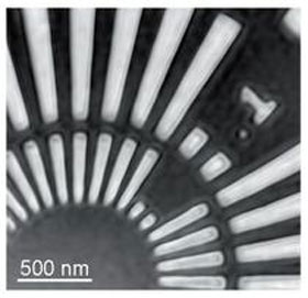 |
| X-ray micrograph of a test pattern made of tantalum measured at PETRA III beamline P06. |
| The scanning X-ray microscope employs a technique known as ptychography. Instead of directly imaging the probe, ptychography combines many diffraction patterns generated when a fine X-ray beam scans the probe. "This way ptychography can overcome the limitations of conventional microscopy regarding the spatial resolution", explains Schroer. |
| The more details on the fringes of the individual diffraction patterns can be recorded, the higher the resulting resolution. Thanks to the extreme brightness of PETRA III the diffraction patterns are exceptionally detailed, resulting in a spatial resolution of ten nanometres, which is at least twice as good as in conventional microscopy. |
| "This is a prime example for the use of a high brilliance source", underlines Schroer. His group demonstrated the capabilities of the new scanning X-ray microscope by imaging a Siemensstern, a star-like pattern with alternating white and black rays, made of the metal tantalum. The technique is suited for a wide range of applications in the nano cosmos as well as in geo- and environmental sciences, and biomedicine. |
| As the resolution of the microscope is in principle only limited by the X-ray density on the sample, an even sharper X-ray vision may become possible in the future. By optimising the focussing X-ray optics a resolution of at least one nanometre should be achievable. |
| Source: DESY |

