| Posted: Jun 18, 2014 |
Microparticles get the whole picture down to the nanoscale |
| (Nanowerk News) Microscopes are conventionally used to image tiny features. However, their resolution is inherently limited by the wavelength of light. This limitation means that they can resolve only structures larger than a few hundred nanometers. Now, Leonid Krivitsky and Boris Luk’yanchuk at the A*STAR Data Storage Institute in Singapore and co-workers have demonstrated an alternative optical approach capable of mapping surfaces at resolutions below 100 nanometers ("Locomotion of microspheres for super-resolution imaging"). |
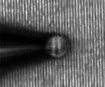 |
| A microsphere connected to the end of a pipette enables sub-diffraction-limit imaging. |
| Diffraction is the tendency for all waves, including light, to spread out when they pass near an object or through a gap. This effect means that optical imaging systems cannot resolve objects smaller than roughly half the wavelength of the illuminating light. Thus, for red light with a wavelength of about 600 nm, the resolution will be approximately 300 nanometers. |
| Luk’yanchuk and his colleagues previously showed that a micrometer-scale transparent bead placed on a surface can circumvent this so-called diffraction limit. They demonstrated that light passing through the bead, when collected by a conventional microscope, can create an image of the surface beneath it with a resolution of 50 nanometers. However, generating a complete two-dimensional map requires scanning the bead across the surface — not easy to perform in a controlled way when the sphere is only 6 micrometers across. “We have now improved this superresolution technique by developing a method to controllably move the imaging microspheres,” says Krivitsky. |
| Krivitsky and his team accomplished such spatial scanning using a tiny pipette with a tip just 1 or 2 micrometers wide. Computer simulations confirmed that the presence of the pipette would not adversely affect the superresolution capability of the microspheres. To fasten the pipette to the bead, they sucked the air out from within its cavity (see image). |
| The team then connected the other end of the pipette to a mechanical stage, which could move in steps as small as 20 nanometers. Importantly, the vacuum inside the pipette created a bond tight enough to ensure that the bead did not disconnect as it was dragged across a surface. The researchers demonstrated the effectiveness of their system by successfully imaging trial samples with features as small as 75 nanometers. |
| While other techniques, such as near-field scanning microscopy, can perform sub-diffraction-limit imaging, they require very expensive systems. “The real advantages of our technique are its simplicity and its price,” says Krivitsky. “The idea could be applied to a variety of superresolution applications such as sample inspection, microfabrication and bioimaging.” |
| Source: A*STAR |

