| Nov 18, 2020 |
Gold nanoparticles turn the spotlight on drug candidates in cells
(Nanowerk News) Successful drug development has a significant impact on people’s quality of life worldwide. Being able to track how molecules get into target cells, and observing what they do when they are inside, is key to identifying the best candidates. Analysis techniques therefore form an important part of the drug discovery process.
|
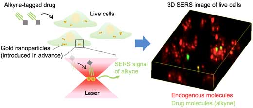 |
| Fig.1: Schematic illustration of alkyne-tagged small-molecule drug detection in live cells using surface-enhanced Raman scattering of gold nanoparticles. (Image: Osaka University)
|
|
Researchers from Osaka University, in collaboration with RIKEN, have reported a Raman microscopy-based approach for visualizing small-molecule drugs that uses gold nanoparticles. The team’s findings are published in ACS Nano ("Quantitative Drug Dynamics Visualized by Alkyne-Tagged Plasmonic-Enhanced Raman Microscopy").
|
|
Small drug molecules are often traced by attaching them to fluorescent probes that are visible when they are irradiated with light. Microscopy can then be used to see these molecules inside cells in real time. However, fluorescent molecules can be bulky, which can affect the way the small molecules behave. Additionally, some fluorescent molecules lose their fluorescence if they are exposed to too much light, making it difficult to see them over the course of long studies.
|
|
One alternative to fluorescent labels is a much smaller tag known as an alkyne, which composed of carbon-carbon triple bonds. The particular arrangement of atoms in alkynes is not found naturally in cells; therefore, they provide a highly specific marker. Furthermore, their small size means that alkynes have minimal effect on the small-molecule behavior. Instead of emitting fluorescence under laser light, alkynes produce what is known as a Raman signal, which can be clearly identified among the cell material signals.
|
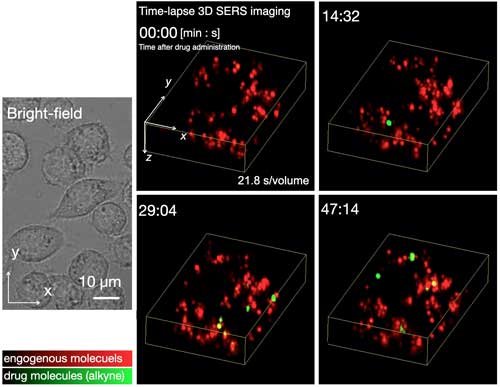 |
| Fig.2: Time-lapse 3D SERS imaging of small-molecule uptake by live cells. Researchers successfully observed that the SERS signals of alkynes were initially detected around 10-15 minutes after drug administration, and the number of the signals gradually increased over time. The drug administration concentration was 20 µM. (Image: Osaka University) (click on image to enlarge)
|
|
However, looking for the Raman signal of alkyne groups is tricky when there aren’t many of them around because of the low efficiency of Raman scattering. The researchers have therefore combined alkyne-tagging with the use of gold nanoparticles. Surface-enhanced Raman scattering (SERS) microscopy can stimulate gold nanoparticles to produce enhanced electric fields that boost the Raman signal of the alkyne groups, making them easier to detect.
|
|
“Our approach is a combination of techniques that have been used for tracking small molecules in live cells,” study lead author Kota Koike explains. “Gold nanoparticles are particularly useful messengers for reporting the presence of alkyne groups because they enhance the alkyne signal, as well as providing a surface that the alkynes like to interact with. The two components therefore come together naturally to generate the enhanced signal.”
|
 |
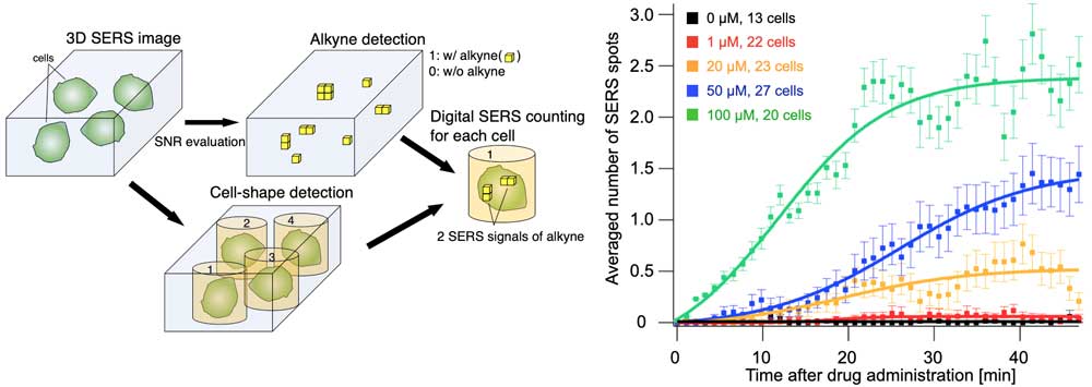
| Fig.3: Quantitative detection of the number of SERS signals of alkynes at the single-cell level (left figure). The number of SERS signals detected per cell at each administration concentration over time (right). Using the quantitative SERS detection method we successfully observed that the uptake speed greatly depended on the drug concentration. (Image: Osaka University) (click on image to enlarge)
|
|
Gold nanoparticles are readily taken up by numerous different types of cells, making the technique broadly applicable. The nanoparticles enter the lysosome compartments inside the cell and then enhance the signal of the alkyne-tagged molecules that subsequently arrive in the lysosomes and interact with them.
|
|
“Our SERS technique has the potential to be used with a variety of different cell types as well as a virtually limitless number of drug candidates,” study corresponding author Katsumasa Fujita explains. “This is particularly exciting for drug discovery where any means of better understanding drug dynamics in real time is extremely valuable for development.”
|