| May 08, 2012 |
Using nanotechnology, researchers discover oldest known blood on 5000 year old mummy
|
|
(Nanowerk News) His DNA has been decoded; samples from his stomach and intestines have allowed us to reconstruct his very last meal. The circumstances of his violent death appear to have been explained. However, what had, at least thus far, eluded the scientists, was identifying any traces of blood in Ötzi, the 5,000 year old glacier mummy. Examination of his aorta had yielded no results. Yet recently, a team of scientists from Italy and Germany, using nanotechnology, succeeded in locating red blood cells in Ötzi's wounds, thereby discovering the oldest traces of blood to have been found anywhere in the world (see paper in Journal of the Royal Society Interface: "Preservation of 5300 year old red blood cells in the Iceman"; open access).
|
 |
|
"Up to now there had been uncertainty about how long blood could survive – let alone what human blood cells from the Chalcolithic period, the Copper Stone Age, might look like." This is how Albert Zink, Head of the Institute for Mummies and the Iceman at the European Academy, Bozen-Bolzano (EURAC) explains the starting point for the investigations which he undertook with Marek Janko and Robert Stark, materials scientists at the Center of Smart Interfaces at Darmstadt Technical University.
|
|
Even in modern forensic medicine it has so far been almost impossible to determine how long a trace of blood had been present at a crime scene. Scientists Zink, Janko and Stark are convinced that the nanotechnological methods which they tested out on Ötzi's blood to analyse the microstructure of blood cells and minute blood clots might possibly lead to a break-through in this area.
|
|
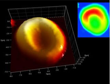
The team of scientists used an atomic force microscope to investigate thin tissue sections from the wound where the arrow entered Ötzi's back and from the laceration on his right hand. This nanotechnology instrument scans the surface of the tissue sections using a very fine probe. As the probe moves over the surface, sensors measure every tiny deflection of the probe, line by line and point by point, building up a three-dimensional image of the surface. What emerged was an image of red blood cells with the classic "doughnut shape", exactly as we find them in healthy people today.
|
|
"To be absolutely sure that we were not dealing with pollen, bacteria or even a negative imprint of a blood cell, but indeed with actual blood cells, we used a second analytical method, the so-called Raman spectroscopy method", report Marek Janko and Robert Stark, who, with Albert Zink, are also members of the Center for NanoSciences in Munich.
|
|
In Raman spectroscopy the tissue sample is illuminated by a laser beam and analysis of the spectrum of the scattered light allows one to identify various molecules. According to the scientists, the images derived from this process corresponded to present-day samples of human blood.
|
|
Whilst examining the wound at the point where the arrow entered the body, the team of scientists also identified fibrin, a protein involved in the clotting of blood. "Because fibrin is present in fresh wounds and then degrades, the theory that Ötzi died some days after he had been injured by the arrow, as had once been mooted, can no longer be upheld," explains Albert Zink.
|

