| Jun 18, 2019 |
Gold nanoparticle clusters for simultaneous photo-thermal imaging and therapy(Nanowerk News) Photo-thermal therapy is a new treatment method for cancer patients that is minimally invasive and can be targeted at a specific region in the body. It uses a photo-thermal agent to absorb and convert light radiation energy into heat to kill the cancer cells (Journal of Physical Chemistry Letters, "Simultaneous Imaging and Selective Photothermal Therapy through Aptamer-Driven Au Nanosphere Clustering"). |
| Gold nanoparticles are excellent photo-thermal agents due to their unique physicochemical properties and high photon-to-heat conversion efficiencies (>99%). However, they have poor light absorption at the near-infrared (NIR) region where tissue transmissivity is optimal. Therefore, a stronger NIR light source has to be used, which could cause unintended collateral damage to surrounding areas. |
| In collaboration with Prof Matthew LANG from Vanderbilt University, the research team led by Prof XU Qing-Hua from the Department of Chemistry, NUS found that gold nanospheres (70 nm in diameter) display enhanced absorption and photo-thermal responses in the NIR region when they aggregate to form clusters. |
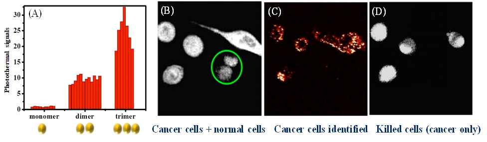 |
| (A) Figure describes the enhanced photo-thermal responses of aggregated gold nanoparticles. (B) Aptamer-modified gold nanospheres displaying high selectivity in specific targeting of human prostate cancer cells over normal human prostate epithelial cells (green circles). (C) Figure shows selective photo-thermal imaging of cancer cells. (D) Figure shows the damaged cancer cells after photo-thermal treatment. (© Journal of Physical Chemistry Letters) (click on image to enlarge) |
| Discrete gold nanospheres are known to be poor near-infrared light absorbers but their absorption ability can be increased by up to 25 times when they form clusters. The enhanced absorption is due to an effect known as plasmon coupling, which occurs when the separation between two particles is less than their individual size. |
| Based on their findings, the researchers have demonstrated photo-thermal imaging and therapy on human prostate cancer cells in a laboratory setting. They designed and built a photo-thermal imaging system that consists of two tightly focused collinear continuous-wave NIR lasers: one beam at 750 nm acts as the heating beam and another beam at 850 nm detects the thermal-induced change in the refractive index. |
| Gold nanospheres modified with aptamers are selectively attached onto the membrane of the human prostate cancer cells to form clusters. This allows the cancer cells to be selectively detected using the system and damaged with high accuracy. |
| Prof Xu said, “Moving forward, the team plans to focus on improving the detection of photo-thermal response through the use of photo-acoustic signals. This will enable more sensitive detection at deeper tissues.” |
| Source: National University of Singapore |
|
Subscribe to a free copy of one of our daily Nanowerk Newsletter Email Digests with a compilation of all of the day's news. |

