| Jul 22, 2019 |
Standard light microscopes get an upgrade to see inside cells(Nanowerk News) Researchers can now see the molecules inside cells using standard light microscopes. Traditionally, light microscopes could only reveal the shape of large structures inside cells, and not the distribution of molecules. |
| The microscope upgrade is an attachable unit that researchers call the “molecular-contrast unit,” which contains an infrared laser light source. No specialized preparation of the cells, for example labeling with fluorescent dyes, is required. |
| The new molecular-contrast unit allows standard light microscopes to "see" where specific molecules are in the cell by searching for the vibration, or heat signal, of the desired molecule. The laser light gives extra energy to the target molecules and causes them to vibrate. Different molecules can be selected by using different wavelengths of infrared laser light. |
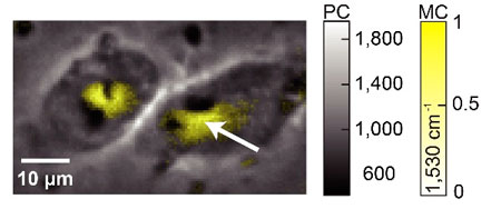 |
| This image of two human cells was made by combining the molecular density information obtained by the molecular-contrast unit (MC, yellow) and the image taken by the conventional phase-contrast portion of a standard light microscope (PC, greyscale). (Image: Takuro Ideguchi, CC-BY) |
| "We believe the concept of upgrading existing, widespread standard optical microscopes to become molecular-sensitive will expand the research capabilities of various end users," said Associate Professor Takuro Ideguchi from the University of Tokyo Institute for Photon Science and Technology. |
| Other methods to see the details of molecules inside cells without adding special dyes already exist, but they require specialized and expensive microscopes or use high-intensity light that can damage the cells. For example, those methods include Raman-scattering and infrared-absorption spectroscopy. |
| "Label-free and damageless molecular microscopic observation, which fulfills an important need in the biomedical field, should be useful for observation of intracellular drug delivery, quality assessment of regenerative cells and tissues, and other essential research functions," said Ideguchi. |
| "Also, since our method translates the local heating of the sample to the molecular contrast, it could also serve as a tool to probe the local thermal parameters within biological cells," he added. |
| So far, researchers have tested their technique using two different types of tiny plastic and silica beads and human cells grown in a dish that were killed and preserved before experiments. |
| "We were pleased to visualize the protein distribution in biological cells. The protein seemed to concentrate around the cells’ nuclei, which could represent the existence of some intracellular structures [involved in the synthesis and transport of proteins], such as the endoplasmic reticulum and Golgi apparatus," said Ideguchi. |
| Collaborators from Osaka University and the Japan Science and Technology Agency also contributed to this research. |
Relevant papers |
| K. Toda, M. Tamamitsu, Y. Nagashima, R. Horisaki, T. Ideguchi, "Molecular contrast on phase-contrast microscope" Scientific Reports, July 18, 2019. |
| K. Toda, M. Tamamitsu, R. Horisaki, and T. Ideguchi, "Phase-contrast microscope with molecular contrast," Conference on Lasers and Electro-Optics/Europe-EQEC, JSII-2.2, Munich, Germany: June 23, 2019 |
| Source: University of Tokyo |
|
Subscribe to a free copy of one of our daily Nanowerk Newsletter Email Digests with a compilation of all of the day's news. |

