| Jan 20, 2021 |
Novel nanomaterial generates powerful beams for enhanced optical imaging
(Nanowerk News) Even the smallest molecule can tell a big story. For instance, observing a single molecule can throw light on underlying biological processes in the human body. In fact, molecular imaging procedures—which are noninvasive and painless—are being used to diagnose and manage the treatment of COVID-19, cancer, heart disease, and other serious health conditions.
|
|
One of the more promising techniques for single molecule imaging is surface-enhanced Raman spectroscopy, or SERS. By focusing a laser beam on the sample, SERS detects changes in molecules based upon how they scatter light, and can identify specific molecules through their unique Raman spectra: a sort of molecular fingerprint. An advantage of SERS is that it is nondestructive and requires minimal sample preparation, as it does not require added chemicals or modifications to take measurements.
|
|
In a study recently published in Advanced Materials ("A Programmable DNA-Silicification-Based Nanocavity for Single-Molecule Plasmonic Sensing"), engineers from Johns Hopkins Whiting School of Engineering describe a novel nanomaterial that enables fast and highly sensitive single molecule detection using SERS. Their invention could pave the way for rapid and more accurate diagnostic testing.
|
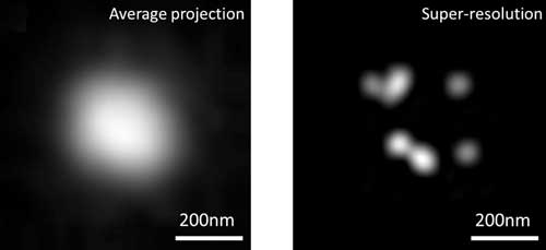 |
| The image on the left (C) shows diffraction-limited imaging that is too blurry to capture plasmonic hotspots required to conduct single-molecule SERS analysis. On the right (D) is super-resolution imaging of the same plasmonic hotspots using DNA-STROBE, which is clear enough to allow for single-molecule SERS analysis.
|
|
To create their new material, called DNA-Silicified Template for Raman Optical Beacon or DNA-STROBE, a team led by Ishan Barman, an associate professor of mechanical engineering, engineered optical cavities of only a few nanometers or fewer. In SERS imaging, these plasmonic cavities "trap" beams of light by converting their electromagnetic radiation into electron waves. The Barman team's tiny plasmonic nanocavities exponentially increase the density of this trapped electromagnetic energy, potentially enabling quantitative biomolecular imaging at ultralow concentrations.
|
|
"The effectiveness of SERS measurements depends on the architecture and reproducibility of the nanoscale probes. If successfully designed and realized, our DNA-STROBE structures offer real-time, single molecule, label-free optical sensing that is almost impossible to achieve with any existing platforms," said Barman, the paper's corresponding author.
|
|
According to Barman, SERS measurements can offer unprecedented insights at the nanoscale, which remains a challenging endeavor for conventional imaging methods. The intensity of the SERS signal depends on the size of the nanoscale gaps, known as "hotspots.". Because these nanocavities confine light energy, the smaller the gaps, the higher the SERS signal. However, nanocavities of this small size are extremely difficult (and expensive) to fabricate in a programmable and reproducible manner, he explained.
|
|
The research team turned to DNA nanotechnology to find an answer. Using DNA as scaffolds, the team built synthetic nanocavities that are the perfect size to become hotspots. But given the elastic nature of DNA, especially its propensity to fold and bend, the size of the formed DNA-STROBE structures could change, potentially weakening the SERS signal. Thus, the team encapsulated the DNA-STROBE structures with a protective ultrathin silica shell to prevent such fluctuations.
|
|
The study reported two significant findings. First, the researchers showed they could fabricate ultra-small nanocavities with well-controlled and large electromagnetic enhancement of the SERS signal. Second, their approach allows for single molecule studies even in biological samples with high molecule concentrations—a roadblock in prior research.
|
|
"We were excited to observe that DNA-STROBE enhanced the Raman signal, and it was strong enough to permit real-time sensing and super-resolution imaging. This will certainly open up new avenues for use of SERS analysis, particularly in sensing and imaging applications where adding contrast agents and dyes is not desirable or practical," said Liang.
|
|
The next step, the researchers say, will be to develop a set of tailored DNA-STROBE-derived analytical tools for a range of applications. For example, the team believes their approach offers a state-of-the-art platform for ultrasensitive detection of circulating cancer biomarkers.
|
|
"With suitable customization, the DNA-STROBE could enable progress in a wide variety of fields ranging from clinical diagnostics and basic biomedical research to environmental sensing and single molecule manipulation," adds Barman.
|

