| Aug 04, 2023 |
Researchers design algorithm to monitor two-photon lithography nanoscale fabrication(Nanowerk News) A new way to monitor two-photon lithography nanoscale fabrication could help improve the accuracy and efficiency of creating 3D engineered tissue scaffolds, according to a new study. Tissue scaffolds mimic the natural extracellular matrices found in the body, which creates a 3D environment ideal for tissue formation. |
| Jieliyue Sun, an engineering Ph.D. student from the lab of Kimani Toussaint, Brown University will present this research at the Optica Imaging Congress. The hybrid meeting will take place 14 – 17 August 2023 in Boston. |
| “Tissue scaffolds are three-dimensional structures that can support the growth and development of cells or tissues for biomedical applications such as tissue engineering, regenerative medicine and drug testing. Cellular behaviors vary with different scaffold geometries at microscale level,” explained Sun. “It is of our interest to investigate those geometric cues in a precisely controlled manner.” |
| Two-photon lithography uses the non-linear phenomenon known as two-photon absorption to produce 3D structures with feature sizes smaller than the diffraction limit. This fabrication approach is well suited for directly writing 3D biomedical scaffolds because it can be used to create high-resolution, well-defined complex 3D microstructures based on computer-aided design (CAD) models. However, assessing the precision of structures fabricated with two-photon lithography has typically required expensive, difficult to implement microscopy methods. |
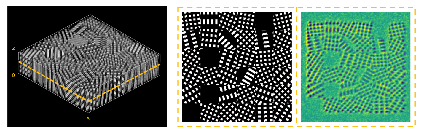 |
| Two-photon fabrication of randomly-oriented fiber scaffolds. Original 3D model (left), sliced input (middle) and fabrication result in the matching layer (right). (Image: Optica) |
| In the new work, the researchers demonstrate a new in-situ monitoring approach that uses adaptive background subtraction for real-time layer-by-layer supervision of two-photon lithography fabrication. It doesn’t require any modifications of the optical system and is relatively simple to implement in most two-photon lithography systems. |
| The new approach uses a monitoring and process control algorithm that enhances the optical sectioning ability of brightfield microscopy in the axial direction. It works by acquiring background images before fabrication begins on each layer and then subtracting the foreground from the adaptive background. This allows the optical contributions from the previously printed layers to be eliminated, revealing single-layer information. |
| The researchers demonstrated the monitoring approach by fabricating a group of synthetic fibers with random orientations, a structure that is similar to that of an arbitrary tissue scaffold. The 3D model was made of 44 sections with a 1-um axial step size. After image processing and cross-correlation calculation, the algorithm was used to determine a quality parameter (q) that indicates the fidelity of the fabrication process. If the q value is below a certain threshold, an error message is generated. |
| Sun added, “With optimized process parameters, we reproduced the input scaffold model with a high geometric fidelity while also revealing the internal features of the architecture. The experiment showed that the new monitoring and process control method improved the quality and efficiency of nanomanufacturing using two-photon lithography. This work paves the way to high-fidelity synthesis of structured tissue scaffolds, and is supported by a grant award from the National Science Foundation’s Division of Civil, Mechanical and Manufacturing Innovation.” |
| Source: Optica (Note: Content may be edited for style and length) |

