| Aug 07, 2023 |
Placing electronics on live cells with nanoscale tattoos
(Nanowerk News) Engineers have developed nanoscale tattoos—dots and wires that adhere to live cells—in a breakthrough that puts researchers one step closer to tracking the health of individual cells.
|
|
The new technology allows for the first time the placement of optical elements or electronics on live cells with tattoo-like arrays that stick on cells while flexing and conforming to the cells’wet and fluid outer structure.
|
|
“If you imagine where this is all going in the future, we would like to have sensors to remotely monitor and control the state of individual cells and the environment surrounding those cells in real time,” said David Gracias, a professor of chemical and biomolecular engineering at Johns Hopkins University who led the development of the technology. “If we had technologies to track the health of isolated cells, we could maybe diagnose and treat diseases much earlier and not wait until the entire organ is damaged.”
|
|
The details are published in Nano Letters ("Toward Single Cell Tattoos: Biotransfer Printing of Lithographic Gold Nanopatterns on Live Cells").
|
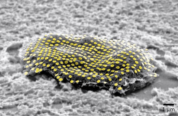 |
| False-colored gold nanodot array on a fibroblast cell. (Image: Kam Sang Kwok and Soo Jin Choi, Gracias Lab/Johns Hopkins University)
|
|
Gracias, who works on developing biosensor technologies that are nontoxic and noninvasive for the body, said the tattoos bridge the gap between living cells or tissue and conventional sensors and electronic materials. They’re essentially like barcodes or QR codes, he said.
|
|
“We're talking about putting something like an electronic tattoo on a living object tens of times smaller than the head of a pin,” Gracias said. “It’s the first step towards attaching sensors and electronics on live cells.”
|
|
The structures were able to stick to soft cells for 16 hours even as the cells moved.
|
|
The researchers built the tattoos in the form of arrays with gold, a material known for its ability to prevent signal loss or distortion in electronic wiring. They attached the arrays to cells that make and sustain tissue in the human body, called fibroblasts. The arrays were then treated with molecular glues and transferred onto the cells using an alginate hydrogel film, a gel-like laminate that can be dissolved after the gold adheres to the cell. The molecular glue on the array bonds to a film secreted by the cells called the extracellular matrix.
|
|
Previous research has demonstrated how to use hydrogels to stick nanotechnology onto human skin and internal animal organs. By showing how to adhere nanowires and nanodots onto single cells, Gracias’ team is addressing the long-standing challenge of making optical sensors and electronics compatible with biological matter at the single cell level.
|
|
“We’ve shown we can attach complex nanopatterns to living cells, while ensuring that the cell doesn’t die,” Gracias said. “It’s a very important result that the cells can live and move with the tattoos because there’s often a significant incompatibility between living cells and the methods engineers use to fabricate electronics.”
|
|
The team’s ability to attach the dots and wires in an array form is also crucial. To use this technology to track bioinformation, researchers must be able to arrange sensors and wiring into specific patterns not unlike how they are arranged in electronic chips.
|
|
“This is an array with specific spacing,” Gracias explained, “not a haphazard bunch of dots.”
|
|
The team plans to try to attach more complex nanocircuits that can stay in place for longer periods. They also want to experiment with different types of cells.
|

