| Nov 01, 2023 |
Scientists expand our understanding of biological nanowires
(Nanowerk News) The movement of electrons across wires is what allows us to use electricity every day. Biological nanowires, microscopic wires made of proteins, have caught researchers’ attention for their ability to carry electrons over long distances.
|
|
In a recent study published in Small ("Long-Range Electron Transport Rates Depend on Wire Dimensions in Cytochrome Nanowires") by the Vermaas lab at the MSU-DOE Plant Research Laboratory, researchers expand our understanding of biological nanowires through the use of computer simulations.
|
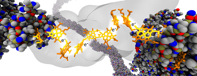 |
| A rendering of a protein nanowire (yellow) cutting through a protein blob (gray) with electron carriers (orange) traveling along it. (Image: Martin Kulke)
|
|
Martin Kulke, first author of the study, accompanied by the Vermaas lab team, created simulations of crystals using data from the real-life experiments in the PRL Kramer lab, where they pointed a light source at a nanocrystal made up of proteins and calculated how fast excited electrons traveled through it. The real question was why electron transfer was getting slower with increasing temperature, which usually accelerates processes at the nanoscale.
|
|
One potential idea was that the distances electrons would need to jump within the nanocrystal might increase with temperature, slowing down how fast they could move through the protein.
|
|
“We simulated these protein nanocrystals at different temperatures to test this idea,” said Josh Vermaas, primary investigator for this study and assistant professor in the Department of Biochemistry and Molecular Biology and at the PRL. “What we found is that the distance changes across different temperatures are not so dramatic on their own.”
|
|
When variables other than temperature were manipulated, the researchers started to see some interesting action from the electrons’ hops within the nanowire. The nanowire protein network was made longer, shorter, thicker and thinner to identify bottlenecks to the electron flow within the nanocrystal.
|
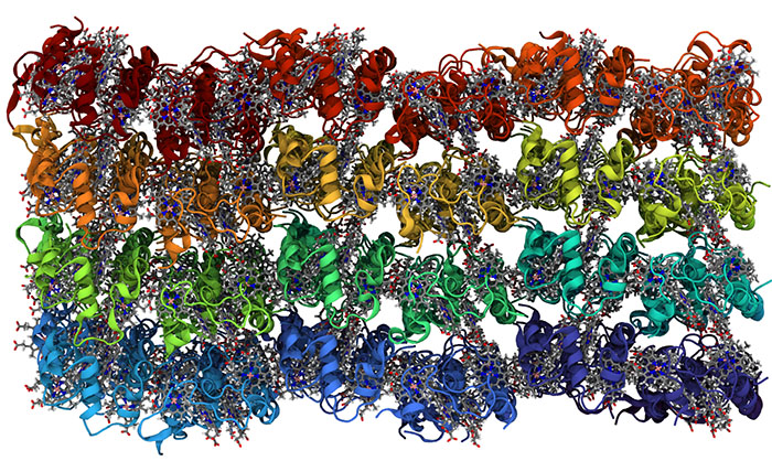 |
| In this representation, each of the 96 proteins in the nanocrystal are a different color. The electrons travel from heme group to heme group inside the protein. The hemes are shown in a stick representation, with gray for the carbons, blue for the nitrogens and a pink iron atom. (Image: Vermaas lab)
|
|
“We found that in biological nanowires, the electron transport is based on the motion of the proteins in the wire,” Kulke said. “What that means is in the end, the longer you make those nanowires, the less electron transport you get through them and the thicker you make them, the more electron transport you get through them.”
|
|
The use of biological nanowires is speculative at the moment, but understanding how they can be constructed to allow for more electron flow is crucial to future endeavors using them to connect biological processes to conventional electronics.
|

