| Jul 16, 2025 |
AI and light-powered nanothermometer maps heat inside living tissue
Researchers have developed an AI-driven nanothermometer that uses invisible light to create 3D temperature maps inside living tissue, enabling safer, noninvasive diagnostics.
(Nanowerk News) A team of researchers from Ca’ Foscari University of Venice and the Universidad Autónoma de Madrid has developed a groundbreaking technique that maps temperature in three dimensions within biological tissue, using invisible light and artificial intelligence.
|
|
The approach, published in Nature Communications ("Luminescence-enabled three-dimensional temperature bioimaging"), could transform how we monitor temperature inside the human body, potentially improving early disease detection and treatment monitoring, without the need for costly or invasive imaging technologies.
|
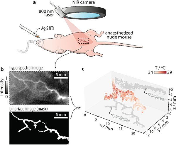 |
| a Representation of an anaesthetized nude mouse to which Ag2S nanothermometers (NTs, 100 μL, 8 mg mL−1) are injected retro-orbitally to obtain a thermal readout of ventral vasculature. b Hyperspectral image (integrated intensity over the 1000–1400 nm range) of the ventral vasculature of the mouse obtained under 808-nm excitation, alongside the binarized image used as a mask to select the pixel to be analyzed are reported. c 3D thermal image of the vasculature obtained from the analysis of the hyperspectral image in (b). The projections of the experimental points along the xy and xz planes are also reported in gray. (Image: reprinted from DOI:10.1038/s41467-025-59681-7, CC BY)
|
|
“We’re turning optical distortions, usually considered a problem, into a source of information,” says Riccardo Marin, associate professor at Ca’ Foscari and one of the lead authors of the study. “With this method, we can detect both how hot a tissue is and how deep it lies beneath the surface.”
|
|
The method relies on luminescent nanothermometers, ultra-small particles made of silver sulfide (Ag₂S) that glow in the near-infrared when stimulated by light. The color and intensity of that glow depend on both the temperature of the particle and the amount of biological tissue the light has to pass through.
|
|
To decode these subtle spectral shifts, the team trained a dual-layer neural network on hundreds of hyperspectral images collected under different conditions. The result is a model that can reconstruct accurate, three-dimensional thermal maps of tissue, even under biologically complex scenarios.
|
|
Proof-of-concept experiments demonstrated the system’s ability to detect temperature gradients in both artificial tissue phantoms and real biological samples. The researchers also succeeded in mapping blood vessels in a living animal, marking the first time that remote, high-resolution 3D thermal imaging has been achieved using light alone.
|
|
While conventional techniques like fMRI or PET scans require costly equipment and specialized training, this new optical method is portable, safer, and significantly less expensive, potentially enabling diagnostics even outside the hospital setting.
|
|
Beyond temperature sensing, the same principles could be adapted to measure other vital parameters such as oxygen concentration and pH, by tailoring the optical properties of the nanoparticles.
|
|
“We believe this is just the beginning,” Erving Ximendes, assistant professor and Ramón y Cajal Fellow at the Universidad Autónoma de Madrid, adds. “Machine learning offers a powerful tool for navigating the complexity of real biological systems—far beyond what traditional models can achieve.”
|
|
The research also highlights the value of international collaboration and talent circulation. The project was initiated during Marin’s time at the Universidad Autónoma de Madrid and involved Anna Romelli, a Ca’ Foscari student on Erasmus mobility. The study now coincides with Marin’s return to Ca’ Foscari, his alma mater, as part of the university’s broader efforts to attract outstanding researchers and strengthen its global research networks.
|

