| Oct 24, 2025 |
AI-driven microscopy is transforming research and sustainable production
AI-enhanced microscopy is reshaping how scientists study particles, improving accuracy, efficiency, and sustainability from early research to industrial use.
(Nanowerk News) A new scientific review (Engineering, "Future Manufacturing with AI-Driven Particle Vision Analysis in the Microscopic World") spotlights particle vision analysis, or PVA, a fast-growing field combining artificial intelligence with microscopic imaging. Researchers say this technology could speed up discovery, improve quality control, and make production more sustainable across industries from nanomaterials to pharmaceuticals.
|
|
Particles lie at the heart of most materials and manufacturing processes. Their tiny shapes, motions, and interactions affect everything from a drug’s stability to a material’s strength. The review explores how AI is helping scientists analyze these particles faster and more accurately, overcoming long-standing challenges in image processing.
|
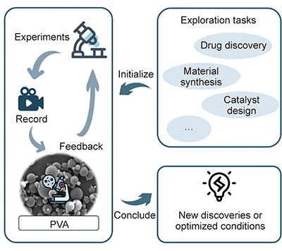 |
| Particle vision analysis links exploration, experiments, and feedback, turning microscopic observations into new discoveries or optimized manufacturing conditions. (Image: Guangyao Chen, Fengqi You)
|
|
The paper maps out how PVA links computer vision techniques—such as object detection, segmentation, tracking, and image enhancement—to real microscopy workflows. It also introduces practical tools and software packages that can plug into laboratory setups or production lines, delivering real-time feedback to fine-tune processes.
|
|
Examples include automated inspection of pharmaceutical vials, on-the-fly particle size checks in factories, and hyperspectral detection of microplastics for rapid environmental testing. Together, these cases show how AI-assisted microscopy can cut waste, boost precision, and shorten development timelines from the nanoscale to mass production.
|
|
Technically, the review highlights breakthroughs that reduce the need for large datasets while improving performance. These include tools such as the Segment Anything Model for image segmentation, CLIP and Grounding DINO for open-vocabulary detection, and fast image recognition systems like YOLO and Mask R-CNN. Imaging advances like ESRGAN and diffusion-based super-resolution further sharpen microscopic detail. One standout example is a zero-shot model that boosts fluorescence microscopy resolution by over 1.5 times without extra training data.
|
|
The authors describe a “discovery-to-optimization” cycle that connects early-stage research with smart manufacturing. AI first helps explore materials or drugs, then analyzes imaging results, and finally feeds that knowledge back to adjust experiments or production conditions—turning microscopic insights into tangible efficiency gains.
|
|
Challenges remain, including particle diversity, noisy data, and the heavy computing needs of high-resolution imaging. The review calls for standardized tools, faster algorithms, and better cross-modality adaptation. Transfer learning and few-shot approaches are seen as promising near-term fixes. To help the field grow, the authors share an open database and code repository, offering a practical starting point for scientists and engineers looking to apply PVA in the lab or on the factory floor.
|

