| Dec 20, 2011 |
Nanoscale growth of cone cells tracked in living human eye |
| (Nanowerk News) Humans see color thanks to cone cells, specialized light-sensing neurons located in the retina along the inner surface of the eyeball. The actual light-sensing section of these cells is called the outer segment, which is made up of a series of stacked discs, each about 30 nanometers (billionths of a meter) thick. This appendage goes through daily changes in length. Scientists believe that a better understanding of how and why the outer segment grows and shrinks will help medical researchers identify potential retinal problems. But the methods usually used to image the living human eye are not sensitive enough to measure these miniscule changes. |
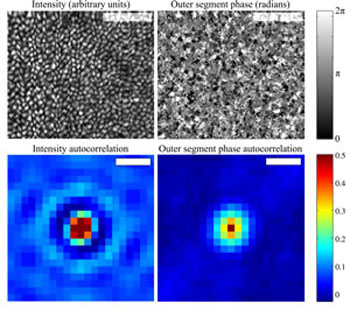 |
| (Upper left) En face projection of the cone mosaic, produced by co-adding intensity from the inner segment outer segment junction (ISOS) and outer segment posterior tip (PT) layers, segmented from a single AO-OCT volume. The bright spots correspond to individual cones. Each cone is ~5 µm in diameter. Scale bar 50 µm. (Upper right) En face projection of the outer segment referenced phase, created by subtracting the phase at ISOS from the phase at PT. Phase correlation is apparent, at a scale similar to that of the intensity projection. Scale bar 50 µm. (Lower left) Autocorrelation of the intensity projection, possessing the stereotypical appearance of a uniformly packed mosaic. The distance between concentric peaks agrees with the predicted cone row spacing. Scale bar 5 µm. (Lower right) Autocorrelation of the referenced phase projection, lacking the concentric rings observed in the intensity autocorrelation. Scale bar 5 µm. The similarity between autocorrelations' central peaks suggests that both intensity and phase are correlated among pixels within the cone, while the dissimilarity between the tails suggests that periodicity exists in the intensity image but not in the phase image. (Image: Ravi Jonnal, Indiana University) |
| Now, vision scientists at Indiana University in Bloomington have come up with a novel way to make the measurements in a living human retina by using information hidden within a commonly used technique called optical coherence tomography (OCT). They discuss their results in the Optical Society's (OSA) open-access journal Biomedical Optics Express ("Phase-sensitive imaging of the outer retina using optical coherence tomography and adaptive optics"). |
| To make an OCT scan of the retina, a beam of light is split into two. One beam scatters off the retina while the other is preserved as a reference. The light waves begin in synch, or in phase, with each other; when the beams are reunited, they are out of phase, due to the scattering beam's interactions with retinal cells. Scientists can use this phase information to procure a precise measurement of a sample's position. But since in this case their samples were attached to live subjects, the researchers had to adapt these typical phase techniques to counteract any movements that the subjects' eyes might insert into the data. |
| Instead of measuring the phase of a single interference pattern, the researchers measured phase differences between patterns originating from two reference points within the retinal cells: the top and bottom of the outer segment. The team used this hidden phase information to measure microscopic changes in hundreds of cones, over a matter of hours, in two test subjects with normal vision. Researchers found they could resolve the changes in length down to about 45 nanometers, which is just slightly longer than the thickness of a single one of the stacked discs that make up the outer segment. The work shows that the outer segments of the cone cells grow at a rate of about 150 nanometers per hour, which is about 30 times faster than the growth rate of a human hair. |
| Source: Optical Society of America |

