| Posted: January 21, 2008 |
New technique will deliver more detailed x-rays |
|
(Nanowerk News) European researchers have developed a way of producing extremely detailed x-ray images using conventional imaging equipment such as that found in hospitals and airports. The new technique produces so-called 'dark-field' images, which could be of use in a number of applications, including medical imaging and security screening.
|
|
Unlike conventional x-rays, dark-field images capture the scattering of the x-rays within the material itself, showing subtle changes in bone, soft tissues and other materials. The image produced is much clearer and more detailed that normal x-rays. In the medical field, the heightened sensitivity of the system means it could be possible to identify minute hairline fractures and measure bone density.
|
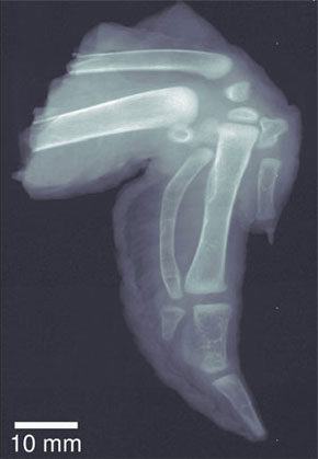 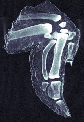 |
| Left: Traditional x-ray image of chicken wing. Right: more detailed darkfield image. (Images: Dr. Pfeiffer, EPFL)
|
|
Furthermore, because cancer and plaque cells scatter radiation differently to normal cells, dark-field x-ray images of soft tissue could show up cancerous tissues and the plaques associated with Alzheimer's. The enhanced sensitivity of the dark-field x-ray can be clearly seen in the dark-field image of a chicken's wing, which is published alongside the article in the journal Nature Materials.
|
|
In security settings, the microcrystalline structure of explosives means they scatter radiation strongly and so show up clearly in dark-field x-rays. In the maritime and aeronautics industries, dark-field x-rays could be used to detect micro-cracks and corrosion on structures such as boat hulls and aeroplane wings.
|
|
However, until now the full potential of these dark-field x-rays has remained largely unexploited because they could only be produced using extremely complex crystal optics and the world's brightest x-ray sources such as synchrotrons.
|
|
In their article, the Danish and Swiss scientists explain how they used nanostructured gratings fitted to conventional x-ray sources to produce dark-field images.
|
|
'Researchers have been working on dark-field x-ray images for many years,' commented Professor Franz Pfeiffer of the Paul Scherrer Institute and École Polytechnique Fédérale de Lausanne in Switzerland.
|
|
'Up until now these images have only been possible using sophisticated crystal optical elements,' added Christian David of the PSI. 'Our new technique uses novel x-ray optical components in the form of nanostructure gratings that permit the use of a broad energy spectrum, including the standard range of energies in traditional x-ray equipment used in hospitals or airports.'
|
|
The scientists now plan to develop an adaptation for use in existing medical equipment. 'When combined with the phase contrast imaging technique that we developed in 2006, we now have the possibility of providing the same range of imaging techniques in broad-spectrum x-ray imaging that we do with visible light,' said Professor Pfeiffer.
|

