| Posted: July 10, 2008 |
Toothpick: New molecular tag IDs bone and tooth minerals |
| (Nanowerk News) Enlisting an army of plant viruses to their cause, materials researchers at the National Institute of Standards and Technology (NIST) have identified a small biomolecule that binds specifically to one of the key crystal structures of the body—the calcium compound that is the basic building block of teeth and bone. With refinements, the researchers say, the new molecule can be a highly discriminating probe for a wide range of diagnostic and therapeutic applications related to bones and teeth. |
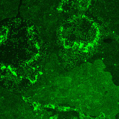 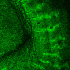 |
| Left: Deposits of hydroxyapatite crystal light up bright green where they've been tagged with a new peptide created at NIST to bond specifically to the compound. The peptide has been linked to a fluorescent stain for imaging. Right: Deposits of hydroxyapatite crystal light up bright green where they've been tagged with a new peptide created at NIST to bond specifically to the compound. The peptide has been linked to a fluorescent stain for imaging. (Images: National Institute of Standards and Technology) |
| Although they have somewhat different mechanical properties, the major structural component of both teeth and bones is a crystalline compound of calcium phosphate called hydroxyapatite. Subtle variations in the way the crystal forms account for the differences. Identifying and monitoring the formation of this particular crystal is of paramount importance to biomedical researchers working on a variety of problems including the remineralization of teeth to repair decay damage, the integration of prosthetic joints and tissue-engineered bone materials for joint and bone replacement, and cell-based therapies to regrow bone tissue. |
| To date, however, there is no specific, practical method to spot the formation of hydroxyapatite in living systems or tissue samples. Materials scientists can identify the crystal structure with high reliability by the pattern it makes scattering X rays, but it’s a complex procedure, requires fairly pure samples and certainly can’t be used on living systems. There are some widely used chemical assays—the von Kossa assay, for example—but these also are destructive tests, and more importantly they really test simply for the presence of the elements calcium or phosphorus. They can’t distinguish, for example, between deposits of amorphous calcium phosphate—a precursor—and the hydroxyapatite crystal. |
| To find a more specific, less destructive probe, the NIST team used a relatively new technique called “phage display” that can rapidly create and screen huge numbers of biomolecules for specific interactions. Phages are a primitive and ubiquitous class of viruses that infect bacteria. Some simple phages can be genetically modified to randomly assemble short sequences of amino acids—small proteins called peptides—on their outer shells as binding sites. An engineered population of phages will synthesize billions of random peptides. If these phages are exposed to the target surface—hydroxyapatite crystal in this case—and then washed off, those left behind are the ones that tend to stick. Cloning the survivors and repeating in several cycles with increasingly stringent conditions eventually isolates a handful of candidate peptides that can be further tested to measure their affinity for the target. |
| As reported in a recent paper ("Identification of a Highly Specific Hydroxyapatite-binding Peptide using Phage Display"), the NIST team used the technique to identify a new peptide that relies both on the chemical composition and the crystal structure of hydroxyapatite to bind to the mineral’s surface. The peptide’s ability to “recognize” the specific structure of hydroxapatite, say the researchers, could be exploited as a nondestructive tag to monitor the progress of bone and tooth mineralization for diagnostic and therapeutic applications. |
| Source: NIST |

