Showing Spotlights 489 - 496 of 546 in category All (newest first):
 Biosensors, which incorporate biological probes coupled to a transducer, have been developed during the last two decades for environmental, industrial, and biomedical diagnostics. The application of nanotechnology to biosensor design and fabrication promises to revolutionize diagnostics and therapy at the molecular and cellular level. The convergence of nanotechnology, biology, and photonics opens the possibility of detecting and manipulating atoms and molecules using a new class of fiberoptic biosensing and imaging nanodevices. These nanoprobes and nanosensors have the potential for a wide variety of medical uses at the cellular level. The potential for monitoring in vivo biological processes within single living cells, e.g. the capacity to sense individual chemical species in specific locations within a cell, will greatly improve our understanding of cellular function, thereby revolutionizing cell biology. Existing nanoprobes have already demonstrated the capability of performing biologically relevant measurements inside single living cells.
Mar 23rd, 2007
 The market for medical implant devices in the U.S. alone is estimated to be $23 billion per year and it is expected to grow by about 10% annually for the next few years. Implantable cardioverter defibrillators, cardiac resynchronization therapy devices, pacemakers, tissue and spinal orthopedic implants, hip replacements, phakic intraocular lenses and cosmetic implants will be among the top sellers. Current medical implants, such as orthopaedic implants and heart valves, are made of titanium and stainless steel alloys, primarily because they are biocompatible. Unfortunately, in many cases these metal alloys with a life time of 10-15 years may wear out within the lifetime of the patient. They also might not achieve the same fit and stability as the original tissue, and in a worst case, the host organism might reject the implant altogether. While available implants can alleviate excruciating pain and allow patients to live more active lives, there often are problems getting bone to attach to the metal devices. Small gaps between natural bone and the implant can increase over time, requiring the need for additional surgery to replace the implant. In the quest to make bone, joint and tooth implants almost as good as nature's own version, scientists are turning to nanotechnology. Researchers have found that the response of host organisms (including at the protein and cellular level) to nanomaterials is different than that observed to conventional materials. While this new field of nanomedical implants is in its very early stage, it holds the promise of novel and improved implant materials.
Mar 22nd, 2007
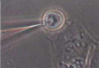
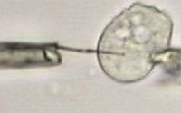 The assembly of nanoparticles along the external or internal surface of carbon nanotubes (CNTs) is of both fundamental and technological interest. Combining unique properties of CNTs and nanoparticles, the nanoparticle/nanotube composite structure attracts a broad range of advanced applications, including nanoelectronics, chemical and biosensors, catalysis and fuel cells. This so-called 'decoration' of CNTs has been used to increase the hydrogen storage capacity, to make nanotubes magnetic, or to grow secondary structures inside the nanotubes to increase the available surface for catalysis. In the case of interior wall decoration of CNTs, the internal cavity of the nanotube often is obstructed and no flow can be achieved or there could be release of the particles in the environment. In the case of exterior wall decorations, the particles enter in direct contact with the environment and may be lost during the nanotube handling. A novel technique of multifunctional nanotubes with controllable amounts of nanoparticles embedded in their walls during the synthesis process solves both problems leaving the CNT bore accessible and keeping the nanoparticles shielded from the environment by the CNT walls. This paves the way to using carbon nanotubes as nanoscale biological probes for sub-cellular investigation.
Mar 16th, 2007
 Can a major component of a catalytic converter or a fullerene derivative lead to an eventual treatment for Parkinson's disease or arthritis? Research to date certainly hints at this possibility. In chemistry, radicals (often referred to as free radicals) are atomic or molecular species with unpaired electrons on an otherwise open shell configuration. These unpaired electrons are usually highly reactive, so radicals are likely to take part in chemical reactions. Radicals play an important role in human physiology but, because of their reactivity, they also can can participate in unwanted side reactions resulting in cell damage. Free radicals damage components of the cells' membranes, proteins or genetic material by "oxidizing" them - the same chemical reaction that causes iron to rust. This is called "oxidative stress". Many forms of cancer are thought to be the result of reactions between free radicals and DNA, resulting in mutations that can adversely affect the cell cycle and potentially lead to malignancy. Oxidative stress is believed to play a role in neurodegenerative diseases such as Alzheimer's and Parkinson's.Some of the symptoms of aging such as arteriosclerosis are also attributed to free-radical induced oxidation of many of the chemicals making up the body. Despite the broad role that oxidative stress plays in human disease, medicine has been limited in its development of treatments that counteract free radical damage and the ensuing burden of oxidative stress. In contrast, in the field of engineering, considerable effort has been developed to counter the effects of oxidative stress at the materials science level. Nanotechnology has provided numerous constructs that reduce oxidative damage in engineering applications with great efficiency. A recent review looks at how these nanoengineering concepts could be applied to biomedical problems, ultimately leading to nanotechnology-based therapeutical treatments for oxidative stress-induced diseases.
Mar 6th, 2007
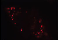 Fluorescent semiconductor nanocrystals (or quantum dots) hold a number of advantageous features including high photobleaching thresholds and broad excitation but narrow emission spectra well suited for multicolor labeling and detection. Unfortunately, most quantum dots are toxic, and hence reduction of cytotoxicity and human toxicity through surface modification plays a pivotal role in their successful application to in vivo labeling, imaging, and diagnosis. Researchers now have demonstrated that nanodiamond particles possess several unique features, including facile surface modification, long-term photostability, and no fluorescence blinking, that makes their detection and long-term tracking in living cells not only possible but practical.
Mar 1st, 2007
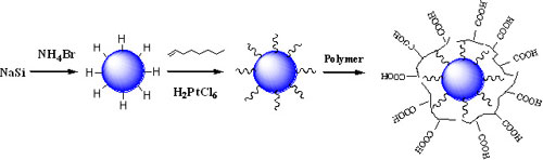 Current medical and biological fluorescent imaging is limited by the use of dye markers, which are not photostable. The dyes can break down under photoexcitation, room light or higher temperatures. The observation of strong visible emission in porous silicon therefore has triggered substantial interest in exploring the synthesis and characterization of silicon nanoparticles. Due to their biocompatibility, high photoluminescence quantum efficiency and stability against photobleaching, silicon nanoparticles are expected to be an ideal candidate for replacing fluorescent dyes in many biological assays and fluorescence imaging techniques. For instance, they have been proposed as better quantum dots for in vivo applications, potentially replacing quantum dots of highly toxic cadmium. Different synthetic and physical methods have been used to prepare silicon nanoparticles. However, the yields of nanoparticles from these methods are very low and an HF (hydrofluoric acid) etching process is often necessary to obtain photoluminescent, hydrogen-terminated silicon nanoparticles. Now, researchers have developed a new solution route for the production of macroscopic amounts of hydrogen terminated silicon nanoparticles without hazardous material handling. This synthesis route is simple and thus offers great opportunity for scaled-up preparation of semiconductor materials.
Feb 19th, 2007
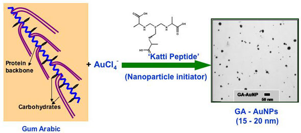 Gold nanoparticles have gained significant prominence in the design and development of nanoscale devices and nanosensors. The ubiquitous place of gold in nanoscience stems from its unique chemical property of serving in the unoxidized state at the nanoparticulate level. In sharp contrast, most of the surfaces of the less-noble metals are susceptible to oxidation to a depth of several nanometers or more, often obliterating the nanoscale properties. The high surface reactivity of gold nanoparticles, coupled with their biocompatible properties, has spawned major interest in the utility of gold nanoparticles for in vivo molecular imaging and therapeutic applications. The core of nanomedicine embodies high surface area and the size relationship of nanoparticles to cellular domains so that individual cells can be targeted for diagnostic imaging or therapy of cancer and other diseases. The development of biocompatible and non-toxic nanoparticles is of paramount importance for their utility in nanomedicine applications. Despite the huge potential for gold nanoparticle-based nanomedicinal products, nontoxic gold nanoparticle constructs and formulations that can be readily administered are still rare. Hypothesizing that the ability of plants to absorb and assimilate metals will provide opportunities to utilize plant extracts as nontoxic vehicles to stabilize and deliver nanoparticles for in vivo nanomedicinal applications, researchers now have used Gum Arabic as a plant-derived, nontoxic construct for stabilizing gold nanoparticles. The development of readily injectable, in vivo stable and non-toxic gold nanoparticulate vectors, especially built from currently accepted human food ingredients, would be pivotal in their many uses (e.g. in vivo sensors, photoactive agents for optical imaging, drug carriers, contrast enhancers in computer tomography, X-ray absorbers).
Feb 12th, 2007
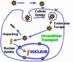 There is currently no cure for Alzheimer's disease and its ultimate cause is still unknown. The disease affects an estimated 4.5 million people in the United States alone. That figure is expected to rise dramatically as the population ages, experts predict. Genetic factors are known to be important in causing the disease, and dominant mutations in different genes have been identified that account for both early onset and late onset Alzheimer's. For a number of years, researchers have been working to alleviate neurodegenerative disorders such as Alzheimer's or Parkinson's disease through gene therapy. In this type of treatment, a gene's DNA is delivered to the neurons in individual cells, allowing them to produce their own therapeutic proteins. Gene therapy typically aims to supplement a defective mutant allele (the location of DNA codings on a chromosome) with a functional one. Currently, the most common carrier vehicles to deliver the therapeutic genes to the patient's target cells are viruses that have been genetically altered to carry normal human DNA. These viruses infect cells, deposit their DNA payloads, and take over the cells' machinery to produce the desirable proteins. One problem with this method is that the human body has developed a very effective immune system that protects it from viral infections. Thanks to advances in nanotechnological fabrication techniques, the development of nonviral nanocarriers for gene delivery has become possible. This is attractive due to the potential for improved safety, reduced ability to provoke an immune response, ease of manufacturing and scale up, and the ability to accommodate larger DNA molecules compared to virus-based delivery tools.
Feb 9th, 2007
 Subscribe to our Nanotechnology Spotlight feed