| Posted: Mar 23, 2015 | |
Nanoparticle shape can drastically enhance MRI signals |
|
| (Nanowerk Spotlight) Magnetic Resonance Imaging (MRI) is a non-invasive medical imaging tool that can provide excellent anatomical information and soft tissue contrast without the need for ionizing radiation or radionuclides. MR images are acquired by exciting protons (in water and fat throughout the body) with a magnetic field and measuring the rate that these protons relax back to their original state. These proton relaxation times vary in different tissues, which results in contrast in the MR image. | |
| Among the lanthanides, gadolinium has been most intensively studied for biomedical applications because, in order to enhance contrast, gadolinium ions (Gd(III)) are often used to shorten the relaxation time of protons (since free lanthanide ions are very toxic, chelating agents are employed to reduce the toxicity). Proton relaxation can be described by relaxivity (r1), which represents how bright the contrast will appear in the MR image. | |
| Typically, in clinical formulations of MRI contrast agents, gram quantities of Gd(III) are needed to achieve sufficiently high contrast for examination. That's why the research imaging community is interested in developing new formulations of contrast agents able to bridge the gap between high contrast imaging of contrast agents dosed at low concentrations. | |
| In new work (ACS Nano, 'Just Accepted Manuscript',"High Relaxivity Gd(III)-DNA Gold Nanostars: Investigation of Shape Effects on Proton Relaxation"), researchers report a new class of gold nanoconjugates that exhibit exceptionally high relaxivities at both low and high field strengths. | |
 |
|
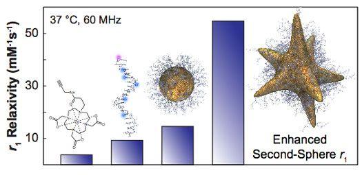
| Gd-DNA@stars achieve r1 relaxivities considerably greater than that observed from their individual components, and much higher than equivalently assembled spherical nanoconstructs. (Image: Odom Group, Northwestern University) | |
| The team, led by Thomas J. Meade, Professor of Chemistry, and Teri W. Odom, Professor of Materials Science and Engineering, both at Northwestern University, designed MRI contrast agents based on nanoparticles that exhibited a remarkably high r1: up to 25 times higher than clinical contrast agents. | |
| "Such a large increase in r1 is significant because it means that lower doses of Gd(III) will be needed to achieve the desired contrast," Odom tells Nanowerk. "We developed nanoconjugate agents by assembling Gd(III)-conjugated DNA onto the surface of gold nanostars, which are anisotropic shaped nanoparticles. Nanoparticle shape, in particular the concave regions of the star, were critical for observing this higher r1. Gold spheres surrounded by the same ligands showed less of an effect. Thus, the nanoscopic spaces created by the negative curvature regions on the gold nanostars were necessary to achieve this enhanced contrast." | |
| The mechanism behind this increase in relaxivity can be attributed to 'second-sphere relaxivity', which means that the Gd(III) ions can coordinate to more water molecules. | |
| "We achieved second-sphere relaxivities that were considerably higher than any previously reported," Meade points out. "Our finding of the importance of nanoparticle shape in manipulating the relaxivity efficiency of surface-bound imaging agents now opens new opportunities in MRI contrast agent development." | |
 By
Michael
Berger
– Michael is author of three books by the Royal Society of Chemistry:
Nano-Society: Pushing the Boundaries of Technology,
Nanotechnology: The Future is Tiny, and
Nanoengineering: The Skills and Tools Making Technology Invisible
Copyright ©
Nanowerk LLC
By
Michael
Berger
– Michael is author of three books by the Royal Society of Chemistry:
Nano-Society: Pushing the Boundaries of Technology,
Nanotechnology: The Future is Tiny, and
Nanoengineering: The Skills and Tools Making Technology Invisible
Copyright ©
Nanowerk LLC
|
|
|
Become a Spotlight guest author! Join our large and growing group of guest contributors. Have you just published a scientific paper or have other exciting developments to share with the nanotechnology community? Here is how to publish on nanowerk.com. |
|