| Posted: Jun 18, 2014 |
Sharper imaging using X-rays
|
|
(Nanowerk News) Physicists at Helmholtz Zentrum Berlin (HZB) have developed a process to generate improved lenses for X-ray microscopy that provide both better resolution and higher throughput. To accomplish this, they fabricate three-dimensional X-ray optics for volume diffraction that consist of on-chip stacked Fresnel zone plates. These three-dimensional nanostructures focus the incident X-rays much more efficiently and enable improved spatial resolution below ten nanometres ("Three-dimensional structured on-chip stacked zone plates for nanoscale X-ray imaging with high efficiency").
|
|
In the future, this kind of novel X-ray optics should be available to users at the BESSY II synchrotron source. Among many applications, the improved resolution permits investigations on ultrastructural features in biological specimens as well as studies on nanostructures in novel battery systems.
|
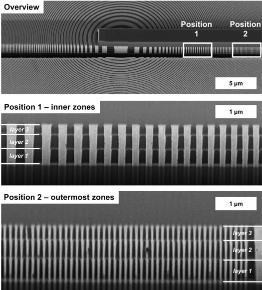 |
| These scanning electron micrographs show how accurately the three Fresnel zone plates were positioned above one another. 3D X-ray optics of this kind allow the resolutions and optical intensities to be considerably improved. (Image: S. Werner/HZB)
|
|
The wavelength of light limits resolution in microscopy. Visible light can resolve structures on the order of a quarter micron, while the considerably shorter wavelength of X-rays can in principle resolve features down to a few nanometres. In addition, X-rays can also penetrate more deeply into specimens, so that internal structures of three-dimensional specimens can be investigated. However, though light in the visible region can be focussed using refractive lenses made of glass, this approach does not work with soft X-rays.
|
|
In order to utilise X-rays for imaging, it is necessary to use Fresnel zone plates, which are made out of concentric rings composed of metals like nickel or gold. These metal rings diffract X-rays so that contributions from the different zones are constructively superposed at the focal point. The result is that Fresnel zone plates act as objective lenses to focus X-rays and can be employed in X-ray microscopes. The achievable spatial resolution depends on the smallest ring width that can be manufactured, which up to now has been about ten nanometres.
|
|
An improvement of spatial resolution to below ten nanometres poses both technological and fundamental physical problems. On the one hand, it is technologically extremely challenging to fabricate periodic zone structures having a ring width of less than ten nanometres and a height of a few hundred nanometres. On the other hand, theoretical calculations indicate that these types of optics with decreasing ring width would be increasingly inefficient and would simply collect too little light. This dilemma can be resolved with the help of volume diffraction. However, the approach requires zone features that simultaneously have an increasing tilt angle and a declining zone height versus radius, i.e. three-dimensional structured X-ray optics.
|
|
|
|
“Theoretically, though, almost 100 per cent of the incident light could be utilised for the image,” explains Dr. Stephan Werner from the Microscopy Research Group at the HZB Institute for Soft Matter and Functional Materials.
|
|
In a first step towards three-dimensional X-ray optics, the experts at HZB have manufactured three layers of Fresnel zone plates nearly perfectly above one another.
|
|
“We have developed a process that enables on-chip stacking of Fresnel zone plates with a precision of less than two nanometres,” says Dr. Gerd Schneider, who heads the Microscopy Research Group.
|
|
The initial measurements demonstrate that this structure captures considerably more light for imaging than conventional Fresnel zone plates.
|
|
“If we are successful in positioning five zone plate layers above one another, which is our next goal, we will be able to utilise a many times higher fraction of the incident X-ray light for imaging than has been available up to now,” says Werner.
|
|
The HZB team is reporting on the development of the novel X-ray optics in the technical journal Nano Research. Users at BESSY II could be soon profiting from this advance as well. X-ray microscopy is an important technique for a wide range of research topics, for example in the life sciences to investigate cell organelles, viruses, and nanoparticles within cells, as well as for materials science and energy research to study novel electrochemical energy storage approaches in situ.
|

