| Posted: Jun 09, 2015 |
3D nano-images reveal 'bicycle spoke' structure of heart cells that may hold key heart attack clues
|
|
(Nanowerk News) Newly released images revealing the ‘bicycle spoke’ structure of a heart cell may hold key clues to reducing damage from a heart attack.
|
|
Research by Dr Ashraf Kitmitto and colleagues, from The University of Manchester’s Institute of Cardiovascular Sciences, provides new information as to why some cells don’t work properly following a heart attack.
|
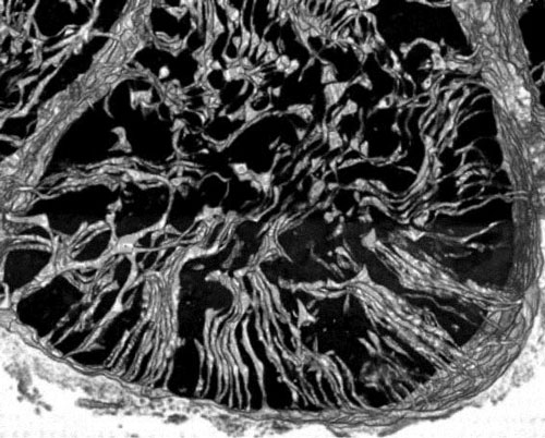 |
| Part of a healthy heart cell with a t-tubule ‘bicycle spoke’ structure.
|
|
Using a novel type of electron microscopy, Dr Kitmitto and team produced 3D images of a healthy heart cell at nanoscopic scale which shows part of their structure is arranged like spokes on a wheel. These ‘spokes’, called T-tubules carry an electrical signal from the outside of the cell to the inside and are necessary for the coordinated transmission of the electrical impulse through the cell to enable the heart cells to contact and enable the heart to pump blood around the body.
|
|
But following a heart attack, the T-tubules are lost in many areas and the electrical signal cannot be carried properly through the cells. The remaining T-tubules also appear to fuse and clump together forming very large, but distorted, ‘super-tubules’.
|
|
This important research, funded by the British Heart Foundation, provides new insights into the structural changes that may contribute towards the development of heart failure and dangerous irregular heartbeats.
|
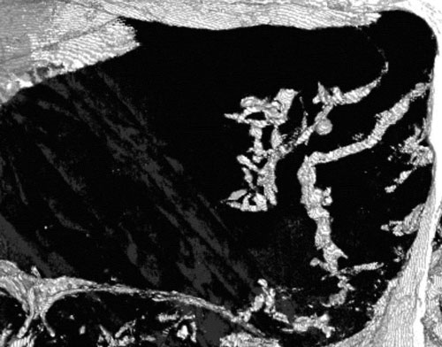 |
| Part of a heart cell where t-tubules have been lost following a heart attack.
|
|
The next step is to find out why this process happens following a heart attack and develop strategies to intervene to stop it from happening, for improved outcomes.
|
|
There are an estimated 550,000 people across the UK living with heart failure, which is when the heart is permanently damaged following a heart attack.
|
|
This research is being presented at the British Cardiovascular Society’s conference in Manchester on Tuesday.
|
|
Dr Kitmitto, whose research was funded by the British Heart Foundation, said: “We’ve made major advances in treating people following a heart attack, so more people are surviving, but the treatments don’t address changes to the structure of the heart.
|
|
“For the first time, we’ve been able to look, in 3D, at the nano-architecture of the cells around the damaged area of the heart and see the changes following a heart attack.
|
|
“The regular pattern of T-tubules – like spokes on a wheel - is really important because it means the whole heart cell can receive the same information and it can contract together.
|
|
“But following a heart attack that regular structure is lost, so some parts of the cell will get the signal and other parts won’t.
|
|
“Now we can see what’s going on, the next step is to find out why and how we can intervene to prevent heart failure development.”
|
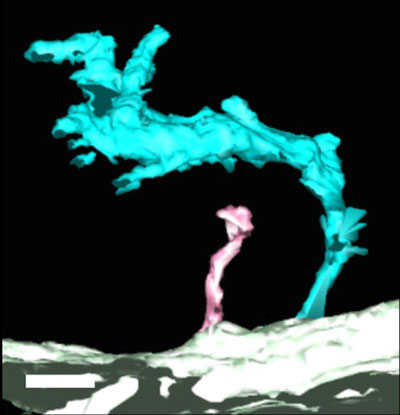 |
| A ‘super-tubule’ (cyan) compared with a healthy t-tubule (pink).
|
|
Dr Mike Knapton, associate medical director at the British Heart Foundation, said: “This interesting research and the beautiful images may hold key clues to reducing the permanent damage caused by a heart attack.
|
|
“Because of the great strides our research has made in treating heart attacks, seven out of ten people now survive.
|
|
“But this means there are an increasing number of people living with damaged heart muscle and heart failure. This research helps us to understand what happens following a heart attack and may lead to treatments to improve the quality of life for heart failure patients in the future.”
|

