| Posted: Sep 16, 2016 |
Silicon fluorescent material developed enabling observations under a bright 'biological optical window'
(Nanowerk News) A research group at the NIMS International Center for Materials Nanoarchitectonics (MANA), led by MANA Principal Investigator Françoise Winnik, a MANA postdoc researcher Sourov Chandra, a research group led by MANA Independent Scientist Naoto Shirahata, and a research group consisting of Professor Yoshinobu Baba and Assistant Professor Takao Yasui, Graduate School of Engineering, Nagoya University, jointly developed a silicon fluorescent material that is very low in toxicity and high in luminescence efficiency, compared to conventional materials.
|
|
Under near-infrared radiation (NIR) at wavelengths of 650 to 1,000 nm—the range known as the “biological optical window”—that is capable of passing through living systems, the joint group succeeded in bioimaging using the new material for the first time in the world (Nanoscale, "Functional double-shelled silicon nanocrystals for two-photon fluorescence cell imaging: spectral evolution and tuning").
|
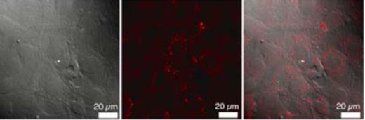 |
| Images of NIH3T3 cells observed under a differential interference microscope (left) and a confocal fluorescence microscope (right). A superimposition of the two images is shown in the middle.
|
|
Fluorescence bioimaging refers to the visualization of cells and other biological tissues that are invisible to the naked eye, by marking them visible with a fluorescent material. The technique enables in vivo observation of the distribution and behavior of living cells in real time.
|
|
Through application of this technique, it may be feasible to observe the behavior of cells and biomolecules linked to pathogenesis and identify the mechanism of disease development. Many of the conventional fluorescent materials emit light when they react to ultraviolet (UV) light or visible light.
|
|
However, because biological components such as hemoglobin and body fluids absorb these types of light, they are not applicable for deep-level observation of biological matters. Some fluorescent materials are reactive to light at wavelengths that fall under a “biological optical window,” but most materials have poor luminescent efficiency, and few others with high luminescent efficiency contain toxic elements such as lead and mercury.
|
|
Using silicon-based particles, the joint group successfully developed a fluorescent material capable of efficiently producing luminescence by reacting to incoming light at wavelengths comparable to a “biological optical window.”
|
|
The use of silicon-based fluorescent materials in bioimaging had been previously studied, and some problems were found such as that they need UV light to exert excitation and efficient luminescence, and that they have low light-emitting efficiencies.
|
| In view of these issues, the joint research group developed a new core-double shell structure in which crystalline silicon nanoparticles, serving as cores, are coated with hydrocarbon groups and a surfactant. Two-photon excitation fluorescence imaging demonstrated that crystalline silicon exhibited efficient photoexcitation when absorbing NIR, and that the hydrocarbon groups in the coating increased emission quantum yield.
|
|
Furthermore, the surfactant coating made the fluorescent material water-soluble. As a result, the new material enabled efficient marking of target biomolecules, and subsequent fluorescent bioimaging of the marked targets using a NIR range of radiation that passes through living systems.
|
|
In future studies, we aim to accomplish fluorescent bioimaging at a deep level using the new silicon fluorescent material we developed in this study.
|

