| Aug 09, 2019 |
Controlling the shape-shifting skeletons of cells(Nanowerk News) You know you have a skeleton, but did you know that your cells have skeletons, too? Cellular skeletons, or cytoskeletons, are shapeshifting networks of tiny protein filaments, enabling cells to propel themselves, carry cargo, and divide. |
| Now, an interdisciplinary team of Caltech researchers has designed a way to study and manipulate the cytoskeleton in test tubes in the lab. Understanding how cells control movement could one day lead to tiny, bioinspired robots for therapeutic applications. The work also contributes to the development of new tools for manipulating fluids on very small scales relevant to molecular biology and chemistry. |
| The work is described in a paper appearing in the journal Nature ("Controlling organization and forces in active matter through optically defined boundaries"). |
| The building blocks of the cellular cytoskeleton are thin, tube-like filaments called microtubules that can form together into three-dimensional scaffolds. Each microtubule is 1,000 times thinner than a human hair and only about 10 micrometers long (about 1,000 times smaller than a common black ant). Along with motor proteins that power movement, these incredibly small structures combine to propel the relatively large cell--like ants steering and powering a car. |
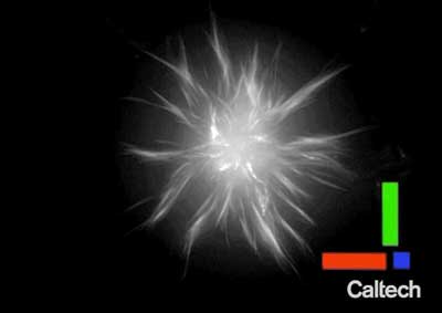 |
| A three-dimensional look at an aster, a structure composed of tiny protein filaments that have been engineered to be controlled with light. (Image: Caltech) |
| In previous studies, researchers have taken these molecules out of the cell and put them into test tubes, where the tubules and motor proteins spontaneously group together to organize themselves into star-shaped structures called asters. How asters in a test tube are related to a cytoskeleton powering cell movement, however, is still unclear. Moreover, the collective microtubule organization demonstrated by aster formation involves interacting forces that are not entirely understood. |
| "What we wanted to know was: how do you get from these spontaneously forming aster structures in the lab, to a cell controlling its movement? And, how can we control these molecules the way a cell does?" says graduate student Tyler Ross, first author on the study. |
| Led by Ross, a team of Caltech researchers explored how to manipulate the component filaments and motor proteins outside of the cell's natural environment. In test tubes, they linked motor proteins to light-activated proteins that are naturally found in plants, so that the tubules would only organize into asters when light was shining on them. In this way, the researchers could control when and where asters would form by projecting different patterns of light, enabling them to develop theories about the physical mechanisms underlying aster formation. |
| Controlling the asters not only allowed for the study of their formation but also enabled the team to build things out of the structures. Ross developed simple procedures of light patterns to place, move, and merge asters of various sizes. The technique offers a way to manipulate structures and study fluid dynamics at a miniscule length scale that is usually difficult to work at; fluids exhibit tricky behaviors at such small volumes. |
| "Generally, it's really difficult to manipulate fluids and structures on this length scale. But this is the scale that we're most interested in for studying cells and chemistry; all of molecular biology works on this scale," says Ross. "Our light-based system allows us to dynamically manipulate our system. We could look through a microscope and say, 'Okay we have enough over here, let's start routing things over there,' and change the light pattern accordingly. We could use aster structures in such a way that they could stir and mix solutions at very small length scales." |
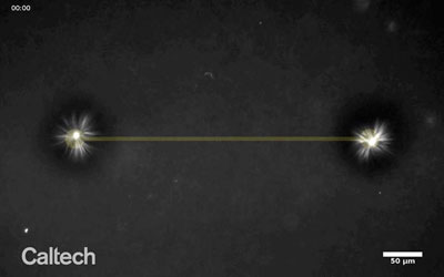 |
| Two asters (white) form and are guided together with beams of light (dark yellow). (Image: Caltech) |
| The research is a collaboration between the laboratories of Matt Thomson, assistant professor of computational biology and Heritage Medical Research Institute Investigator, and Rob Phillips, Fred and Nancy Morris Professor of Biophysics, Biology, and Physics. This collaboration, notes Thomson, enabled pivotal breakthroughs in the project, which Ross had begun in Thomson's laboratory at UC San Francisco (UCSF) before the two came to Caltech in 2017. At Caltech, the pair teamed up with Heun Jin Lee, a staff scientist with extensive expertise in optics, to develop a specialized microscope with which they could view aster formation and direct precise patterns of light. |
| "This has been one of the great collaborations I've seen in my career," says Thomson. "This story really speaks to the community, how you can do work across different fields and people will support and cultivate it. We had feedback from people who work in DNA nanotechnology and people who work in chemical engineering and fluid dynamics." |
| Source: California Institute of Technology |
|
Subscribe to a free copy of one of our daily Nanowerk Newsletter Email Digests with a compilation of all of the day's news. |

