| Nov 02, 2012 |
Giving fluorescence microscopy new power to study cellular transport
|
|
(Nanowerk News) The ability of fluorescence microscopy to study labeled structures like cells has now been empowered to deliver greater spatial and temporal resolutions that were not possible before, thanks to a new method developed by Beckman Institute faculty member Gabriel Popescu and Ru Wang from his research group. Using this method, the researchers were able to study the critical process of cell transport dynamics at multiple spatial and temporal scales and reveal, for the first time, properties of diffusive and directed motion transport in living cells.
|
|
Popescu leads the Quantitative Light Imaging Laboratory at Beckman, while Wang of the lab is first author on the paper reporting the method in Physical Review Letters ("Dispersion-Relation Fluorescence Spectroscopy"). The new approach, called dispersion-relation fluorescence spectroscopy (DFS), labels molecules of interest with a fluorophore whose motion, the researchers write, “gives rise to spontaneous fluorescence intensity fluctuations that are analyzed to quantify the governing mass transport dynamics. These data are characterized by the effective dispersion relation.”
|
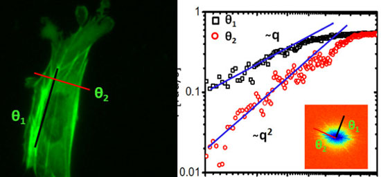 |
| Dispersion-relation fluorescence spectroscopy of mouse embryonic fibroblast (MEF): a) fluorescence image showing a cell whose actin was labeled with GFP. b) Dispersion curve measured for the cell in a. The black and red lines indicate directed motion (along the actin filaments) and diffusion (perpendicular to the actin filaments). Inset shows the dispersion map. (Image: Gabriel Popescu)
|
|
That ability to study the directed and diffusive transport characteristics of cellular dispersion through a wide range of temporal and spatial scales is more comprehensive than using just fluorescence microscopy. It provides more information than existing methods, such as fluorescence correlation spectroscopy (FCS), which is widely used for studying molecular transport and diffusion coefficients at a fixed spatial scale.
|
|
This study used DFS to focus on the cell cytoskeleton subunit actin and found that “the fluorescently labeled actin cytoskeleton exhibits active transport motion along a direction parallel to the fibers and diffusive on the perpendicular direction.” Those results, the researchers said, describe at what scale and when directed versus diffusive motion is taking place in the cell.
|
|
“So for the first time we think we’re able to tell those apart and the spatial scales at which each is dominant,” Popescu said.
|
|
“Some traditional methods are good at measuring local transport and some are good at measuring the larger scales,” Wang said. “Our method gives a fuller view of what happens inside the cell, to the patterns of traffic. So we can look at both the local scale and at larger scales, and ask at which scale the motion transitions from random to directed motion.”
|
|
Popescu said the multiplicity of scales the method offers over techniques like fluorescence correlated spectroscopy is key.
|
|
“It’s like looking from the moon at a highway system in North America and you’re trying to understand the traffic,” Popescu said. “There are so many paths and some cars are moving fast, some slow, some over short distances, some over large distances, and all of these things are happening at the same time. We are actually able to break that information down to these simple pieces that seem to represent a universal behavior for all the cells we measured.
|
|
“With local measurements, it’s actually difficult to measure all these complexities because you only have one point of measurement. That’s why we tried to search for a better way that also uses the spatial information of that traffic. I think we now have solved it.”
|
|
Such knowledge would be valuable for researchers interested in the basic science of cellular dynamics, as well as those working in biomedical research, such as in analysis of a drug’s effect on the body. This technique can be used with current fluorescence microscopy methods.
|
|
“I think that the beauty of this method is that you can use a commercial fluorescent microscope that is found everywhere to collect and analyze data in a very simple way,” Wang said. “You don’t need complicated expertise. Everyone can use it.”
|
|
The method relies on taking time-resolved sequential data from fluorescent spectroscopic microscopy images and transforming them using the Fourier transform. This computational method enables easier understanding of the image data, providing a different representation of the image. Taking advantage of the respective frequency domains of patterns in the data, as this method does, is especially useful for trying to understand cellular dynamics like transport.
|
|
“It turns out the laws of physics are actually best described in the frequency domain,” Popescu said. “The dispersion relation in all branches of physics connects spatial scales with temporal scales. For example, as things get smaller in space, in length if you like, they tend to move faster. A fly will move faster than an elephant.
|
|
“This dispersion relation tells you how much faster. If I make something twice as small, is it going to move twice as fast, or four times or eight times? This relationship basically tells you everything about that dynamic phenomenon. So for the first time we saw this universal transport behavior in a living system: a clear combination of diffusive transport, like Brownian motion, and directed, deterministic transport. As a general trend, we found that diffusion is dominant at short scales and directed transport at large distances.”
|
|
They have also used the method to study neurons in work with the Center for Emergent Behaviors of Integrated Cellular Systems (EBICS) at Illinois, a multi-university project aimed at building living, multi-cellular machines that address real-world problems. The revelations regarding directed versus diffusive transport could be especially useful in reaching that goal.
|
|
“The fact that we can tell where the deterministic and the random transport appears is actually very relevant for looking at cells as a machine,” Popescu said. “What makes a cell machine is actually this directed component because you cannot predict with accuracy Brownian motion, but you can predict this directed motion.”
|
|
The group collaborated with Peter Wang’s lab in that research. Popescu said the collaboration with Wang and EBICS is just one example of the potential of DFS to be useful in many areas.
|
|
“We are measuring neuron networks. We’ve already shared these results with the Center and they are very excited,” Popescu said. “This is a very broadly applicable method.”
|

