| Jul 22, 2013 |
Putting the squeeze on cells to deliver
|
|
(Nanowerk News) Imagine being able to redirect powerful immune cells to fight cancer. How about reprogramming a diabetic’s skin cell into a cell that could manufacture the insulin their pancreas no longer produces? Could we dial down the production of fat cells in obese adolescents? These are major health problems and medical challenges that may be more achievable with a new fundamental technology that gets vital control molecules into cells faster, safer, and more effectively.
|
|
NIBIB-funded engineers at the Massachusetts Institute of Technology (MIT) have developed a rapid and highly efficient system for transferring large molecules, nanoparticles, and other agents into living cells, providing new avenues for disease research and treatment. Cells carrying these “transferred molecules”– the intended therapy - can be used in many ways, including therapeutic and diagnostic interventions in patients and experimental therapies in animal models of disease. The technique offers a powerful tool for probing how cells and their molecular components work by studying how transferred molecules affect a cell’s behavior and functions.
|
|
The system uses controlled mechanical force (relatively gentle squeezing) that is non-toxic to cells, unlike other methods that use viruses, chemicals or electric shock, which can kill cells and damage the transferred molecules. In addition, the new device is “high throughput,” which means it works rapidly, treating a remarkable 20,000-100,000 cells per second.
|
|
The speedy transfer of therapeutic molecules into cells with minimal cell damage and death allows millions of cells to be treated in a very short period of time. This is important because usually, large numbers of treated cells are needed to achieve diagnostic and therapeutic effects.
|
|
The system was developed through a collaboration between the laboratories of Robert Langer and Klavs Jensen, both at MIT. The work is published in the February 5 edition of the Proceedings of the National Academies of Science ("A vector-free microfluidic platform for intracellular delivery").
|
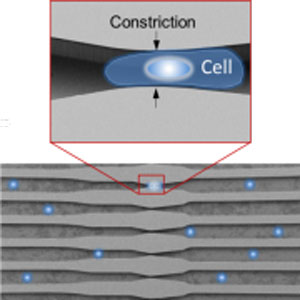 |
| The cell membrane is disrupted when the cells (blue dots) are forced through constrictions in the channels of the microchip.
|
|
How it works
|
|
The device, known as a microfluidic delivery platform, is made up of channels etched into a silicon microchip through which cells initially can flow freely. However, as the cells move through the device --like an inner tube along a water slide-- the channel width narrows until a cell finds itself in a tight spot -- forced to fit through a space that is narrower than the cell. The supple cell membrane allows the cell to squeeze through the constriction. However, the forced, rapid change in cell shape creates temporary holes in the cell membrane, without permanently damaging or killing the cell.
|
|
While the cell membrane is temporarily disrupted, the molecules to be delivered pass through the holes in the membrane and enter the cell. As the cell rebounds to its normal shape, the holes in the membrane close; the cell is loaded successfully. Virtually any type of molecule can be delivered into large numbers of any type of cell.
|
|
New technique expands experimental and therapeutic possibilities
|
|
The new system has distinct advantages over those currently in use. For example, a common technique known as electroporation cannot successfully deliver nanoparticles because they get damaged or inactivated by the electric pulse that is applied to disrupt the cell membrane. Two such nanoparticles are quantum dots and gold nanoparticles, which can be used to track cells inside the body because their electrical properties make them highly visible using biological imaging techniques. Using the new MIT technique, which disrupts the cell membrane by mechanical force rather than an electrical pulse, the team successfully transferred these nanoparticles without damaging their electrical properties. Therefore, the new technique allows researchers to load these electrically sensitive nanoparticles into cells and follow them through the body to diagnose disease and monitor treatments.
|
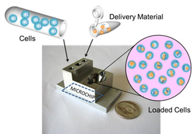 |
| Cells and the delivery material are added separately and flow together through the small device that houses the microchip. The pink circle depicts cells successfully loaded with the delivery material.
|
|
Another significant advantage of the relatively gentle, yet highly effective technique is the ability to transfer molecules into fragile cells that do not survive the current methods. One such cell type is skin cells taken directly from an individual. Using the new mechanical force system the team successfully transferred a set of proteins into freshly obtained human skin cells, where the proteins acted to transform the skin cells into stem cells. Stem cells are an “all purpose” type of cell that scientists are eagerly working with to develop new regenerative therapies. The ability to easily make stem cells from an individual’s skin cells, using this new technique, is a significant step that promises to accelerate the development of stem cell based therapies to regenerate diseased or damaged tissues.
|
|
Some of the most exciting uses for this new system are likely to take the form of novel therapies. Armon Sharei, a graduate student in the Jensen laboratory and one of the lead developers of the technique, described a therapeutic application that the researchers are particularly excited about: “Our big push today is in the field of immunology. Immune cells are very resistant to traditional transfer techniques, yet they hold enormous therapeutic potential. In close collaboration with other laboratories, we hope to use this technology to harness the power of the patient’s own immune system to combat complex immune disorders that currently have no effective treatments.”
|
|
The project was supported partially by an American Recovery and Reinvestment Act (ARRA) NIH Challenge Grant. The special two-year grants supported research on Challenge Topics that addressed specific scientific and health research challenges in biomedical and behavioral research. Dr. Rosemarie Hunziker, the NIBIB Program Director for Tissue Engineering and Regenerative Medicine elaborates: “The goal of the ARRA awards was to support studies that could produce important innovations within a short time frame. We certainly achieved that here. This deceptively simple new way to control cell behavior offers exciting promise for studies of basic cell biology as well as enabling cell-based therapies previously only envisioned. Now that the basic principle has been established here, numerous novel applications can be pursued by diverse teams of scientists and engineers. It’s what we do at NIBIB--enable technologies capable of making a profound difference in medical care and the lives of patients.”
|

