| Aug 06, 2013 |
New microchip sorts white blood cells from whole blood (w/video)
|
|
(Nanowerk News) Early in 2012, MIT scientists reported on the development of a postage stamp-sized microchip capable of sorting cells through a technique, known as cell rolling, that mimics a natural mechanism in the body. The device successfully separated leukemia cells from cell cultures — but could not extract cells directly from blood.
|
|
Now the group has developed a new microchip that can quickly separate white blood cells from samples of whole blood, eliminating any preliminary processing steps — which can be difficult to integrate into point-of-care medical devices. The hope, the researchers say, is to integrate the microchip into a portable diagnostic device that may be used to directly analyze patient blood samples for signs of inflammatory disease such as sepsis — particularly in regions of developing countries where diagnostic lab equipment is not readily available.
|
|
In their experiments, the scientists pumped tiny volumes of blood through the microchip and recovered a highly pure stream of white blood cells, virtually devoid of other blood components such as platelets and red blood cells. What’s more, the team found that the sorted cells were undamaged and functional, potentially enabling clinicians not only to obtain a white blood cell count, but also to use the cells to perform further genetic or clinical tests.
|
|
Rohit Karnik, an associate professor of mechanical engineering at MIT, says the key to recovering such pure, functional cells lies in the microchip’s adaption of the body’s natural process of cell rolling.
|
|
“We believe that because we’re using a very biomimetic process, the cells are happier,” Karnik says. “It’s a more gentle process, and the cells are functionally viable.”
|
|
Karnik and MIT graduate student Suman Bose, along with Jeffrey Karp at Brigham and Women’s Hospital and five other colleagues, published their results in the journal Scientific Reports ("Affinity flow fractionation of cells via transient interactions with asymmetric molecular patterns").
|
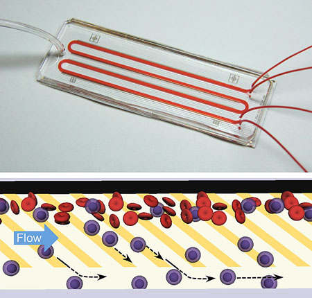 |
| A microfluidic chip, like the one shown above, directly separates neutrophils from blood with ultrahigh purity and high efficiency without the need for cumbersome sample preparation. The chip works by mimicking the physiological process of 'cell rolling' where patterns of adhesive molecules are used to draw out neutrophils (blue) from a stream of blood (red) into a parallel buffer stream as shown in the bottom panel. (Images: Suman Bose and Rohit Karnik)
|
|
Getting a picture of cell rolling
|
|
Normally, the body’s protective white blood cells circulate through the bloodstream, patrolling for signs of pathogens. When one region of the body becomes infected or inflamed, cells lining the blood vessels in that region present certain sticky molecules that gently grab white blood cells from the bloodstream, “rolling” the cells along the vessel wall until they reach the afflicted area.
|
|
The sticky molecules act as cell traffickers, directing particular cells to areas of the body where they’re needed. One of the more common cell traffickers is P-selectin, a molecule that lightly binds the white blood cells called neutrophils.
|
|
In their 2012 study, the researchers first etched the chip with a pattern of ridges and troughs, and lined the ridges with P-selectin. As they pumped a cell culture through the chip, the sticky ridges pulled leukemia cells out of solution, flipping them into trenches that flowed into a separate receptacle. Although they recovered the target cells at high throughput, suitable for laboratory cell separations, the researchers observed that the pattern of grooves caused unwanted mixing of fluid, making it unsuitable for processing blood.
|
|
For blood samples, Karnik and Bose worked in parallel on a different microchip design. Instead of grooves, the team etched a pattern of microscopically thin gold stripes, diagonal to the flow of fluid through the chip. The researchers then attached molecules of P-selectin along each gold stripe.
|
|
Before flowing blood through the chip, the team first tested the design with a culture of two types of leukemia cells. Through video imaging, they observed that one cell type was drawn out of the stream, attaching and rolling briefly along a sticky gold stripe before jumping to a neighboring line. Eventually, after a series of jumps and rolls, the cells rolled the rest of the way along a stripe, then into a separate gutter, or channel, where they flowed into a collector.
|
|
|
|
Neutrophils, a type of white blood cell, follow patterns of weak adhesive molecules (dark lines) and are separated from a stream of blood flowing in a microfluidic channel. (Video: Suman Bose and Rohit Karnik)
|
|
Karnik and Bose measured the interactions between target cells and the chip’s pattern of stripes, and developed a mathematical model to describe the optimal pattern of cell rolling, depending on several factors such as the angle and length of each stripe.
|
|
“We are able to get a good picture of the separation process, and this should be useful in guiding future devices of this type,” Karnik says.
|
|
At the patient’s bedside
|
|
Using the model, the group optimized the chip’s pattern of stripes, then flowed tiny volumes of blood through an inlet. They found they were able to continuously recover a stream of white blood cells with more than 99 percent purity — a significant result, as there are normally 1,000 red blood cells for every white blood cell circulating in the bloodstream.
|
|
In tests of viability, Karnik and Bose found the recovered white blood cells were able to successfully eat up bacteria, signaling that the cells were indeed functional.
|
|
Going a step further, the group tested a potential clinical application: diagnosing sepsis, a condition in which infection spreads through the bloodstream, causing widespread infection and eventual organ failure. The condition is difficult to quickly diagnose, especially in resource-limited settings.
|
|
To mimic conditions of sepsis in blood, the group added cytokines and bacterial membrane components — inflammatory agents typically present during sepsis — to whole blood. It is known that in the presence of such agents, white blood cells lose their ability to bind with P-selectin during sepsis.
|
|
Just as they expected, when the researchers flowed only a few microliters of sepsis-mimicking blood through the microchip, they observed a sharp decline in the number of recovered white blood cells. The result, Bose says, is a promising step toward future diagnostics for inflammatory conditions like sepsis, particularly for babies.
|
|
“It’s more difficult for babies, because for them you can’t get more blood samples because they have so little blood,” Bose says. “This is just a teaser into a new direction of care, and where the work can go in terms of diagnosis of inflammation right at the patient’s bedside.”
|
|
Xuonhong Cheng, an assistant professor of materials science and engineering at Lehigh University, says the purity of samples separated by the group’s device is comparable to that of conventional cell-sorting techniques, “but with much less manual handling and contamination of red blood cell debris.”
|
|
Cheng, who was not involved in the research, says the cell-rolling technique “is a very promising approach to automate blood sample preparation in clinical applications [such as] infection and inflammation assays at point-of-need environments.”
|

