| Posted: Jul 13, 2007 | |
Reduced quantum dot toxicity through 'jelly dots' |
|
| (Nanowerk Spotlight) A quantum dot (QD), also called a nanocrystal, is a semiconductor nanostructure that can be as small as 2 to 10 nm. The usefulness of quantum dots comes from their peak emission frequency's extreme sensitivity - quantum mechanical in nature - to both the dot's size and composition. QDs have been touted as possible replacements for organic dyes in the imaging of biological systems, due to their excellent fluorescent properties, good chemical stability, broad excitation ranges and high photobleaching thresholds. However, the main drawback of QDs is their toxicity and therefore their application is problematic. If this toxicity problem could be addressed, QDs may one day be safely utilized in many areas. For instance, cadmium telluride (CdTe - which is toxic) QD based nanocomposites can be used as fluorescent probes for biological imaging, they can also be utilized to monitor targeted drug delivery and for controlled modification of structural and functional properties of intracellular components. Scientists in Ireland have been using gelatin during the production of CdTe QDs thereby reducing the toxicity of the particles. Their approach could be useful for the development of other nanoparticle composites with low toxicity as well. | |
| Nanotoxicity is now one of the most topical subject that influences development and applications of nanotechnology and nanomaterials. More and more, nanomaterials are becoming part of our daily lives with their applications raging from sunscreens to lightweight sports equipment. One important, and very promising, area of nanotechnology is nanomedicine. It is difficult to find fault with a technology that promises to cure cancer almost before it starts and prevent the spread of AIDS and other infectious diseases. Scientists around the globe are searching for ways to exploit nanoparticles for the benefit of human health. The medical advancements that may be possible through nanotechnology range from diagnostic to therapeutic, and everything in between. | |
| Quantum dots in particular have taken the step from pure demonstration experiments to real applications in imaging. In recent years, scientists have discovered that these nanocrystals can enable researchers to study cell processes at the level of a single molecule and may significantly improve the diagnosis and treatment cancers. Fluorescent semiconductor quantum dots are proving to be extremely beneficial for medical applications, such as high-resolution cellular imaging. Quantum dots could revolutionize medicine, unfortunately, most are toxic. | |
| The aggregation, stability, and degradation of QDs in an intracellular environment is of major concern and understanding their biological behavior is of paramount importance. Recently, several investigations have concluded that QD toxicity is highly dependent not only on the core and surface functionality but also on their size. It also was found that coating the QDs with an epitaxial shell can increase biocompatibility and reduce leaching of the toxic ions from the metalloid core. | |
| "Previously, work has centered on the determination of toxicity related phenomena with regards to the use of quantum dots (QDs) for imaging cellular systems" Dr. Yurii Gun'ko explains to Nanowerk. "This has revolved around the analysis of toxicity due to leeching of toxic elements from the core, the interaction of the capping molecules with the medium and the presence of a nanomaterial in a biological environment. Our recent work strives to reduce this inherent toxicity by adding gelatin as a co-capping agent to reduce the impact that the QDs have on the cells." | |
| Gun'ko, a lecturer in Inorganic Chemistry at Trinity College Dublin, and a principal investigator at CRANN, Ireland's Centre for Research on Adaptive Nanostructures and Nanodevices, together with collaborators from CRANN, was able to demonstrate the reduced toxicity of the CdTe QD–gelatin nanocomposites and their potential as bioimaging agents. The researchers reported their work in a recent article in Small ("'Jelly Dots': Synthesis and Cytotoxicity Studies of CdTe Quantum Dot–Gelatin Nanocomposites"). | |
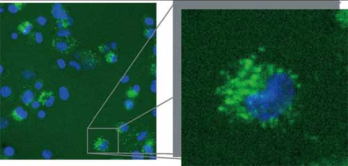 |
Live-cell Cellomics image illustrating the uptake and biocompatibility of the gelatin–QDs. The QDs readily pass through the cellular membrane; however, no nuclear penetration is observed. (Reprinted with permission from Wiley) |
| "We have utilized gelatin in situ during the production of CdTe QDs" says Gun'ko. "This is the main novelty in our approach. The gelatin polymer interacts with the cadmium precursors altering the growth dynamic of the system and increasing the luminescent properties of the QDs. It also helps to reduce the toxic effects induced by the QDs once incubated in the presence of THP-1 macrophage cells." | |
| Some recent publications have highlighted the use of gelatin nanoparticles (for instance: "In vitro uptake of gelatin nanoparticles by murine dendritic cells and their intracellular localisation"), and also their encapsulation in a silica shell, as new biopolymer-based nanocomposites in an effort to generate a biocompatible nanoscale probing system. In their paper, the scientists in Ireland report for the first time the use of gelatin in situ for the production of CdTe QD–gelatin nanocomposites. | |
| "We have examined the biocompatibility and toxicity of these QD systems in parallel to controls of unmodified cells and those containing only free cadmium, by using a Cellomics high-content analysis approach" says Gun'ko. "The gelatin–QD composites readily pass through the cell membrane and illuminate the cytoskeleton of the THP-1 macrophage cells. In comparison to the original thioglycolic acid stabilized QDs, the gelatin–QDs display much lower rates of toxicity (assessed through decreased cell permeability and an aversion to increased lysosomal pH), which are comparable to those of control samples. The free cadmium demonstrates the highest responses for both of these tests and quickly induces cellular apoptosis. | |
| Gun'ko and his colleagues believe this approach can be used for the development of other nanoparticle composites with low toxicity. These composites may have a range of potential biomedical applications including diagnostic recognition and treatment of many diseases. | |
| In the long term this research might also enable scientists to understand the mechanistic pathways of penetration by very small toxic particles (for example cancerogenous dust nanoparticles) and viruses into immune systems cells and help in diagnostic recognition and treatment of cancer, HIV and many other diseases. | |
| In a next step the researchers will try to develop biocompatible quantum dots possessing specific functionalities for targeted intracellular localization and investigation of the mechanistic pathways. This would then allow them to explore potential applications of quantum dots for monitoring of drug delivery and examination of various intracellular processes. | |
| "There are a lot of potential challenges for this research" says Gun'ko. "We believe that anticipated challenges and technical problems will mostly be related to the physiological stability, toxicity and biocompatibility quantum dot based composites." | |
 By
Michael
Berger
– Michael is author of three books by the Royal Society of Chemistry:
Nano-Society: Pushing the Boundaries of Technology,
Nanotechnology: The Future is Tiny, and
Nanoengineering: The Skills and Tools Making Technology Invisible
Copyright ©
Nanowerk LLC
By
Michael
Berger
– Michael is author of three books by the Royal Society of Chemistry:
Nano-Society: Pushing the Boundaries of Technology,
Nanotechnology: The Future is Tiny, and
Nanoengineering: The Skills and Tools Making Technology Invisible
Copyright ©
Nanowerk LLC
|
Become a Spotlight guest author! Join our large and growing group of guest contributors. Have you just published a scientific paper or have other exciting developments to share with the nanotechnology community? Here is how to publish on nanowerk.com.