| Posted: Nov 13, 2014 | |
Graphene electrodes for simultaneous electrophysiology and neuroimaging |
|
| (Nanowerk Spotlight) Studying the complex wiring of neural circuits and identifying the details of how individual neural circuits operate in epilepsy and other neurological disorders requires real-time observation of their locations, firing patterns, and other factors. These observations depend on high-resolution optical imaging and electrophysiological recording. | |
| "Metal microelectrode arrays commonly used for recording neural activity cannot be used for such purposes, as they block the field of view, generate optical shadows and are prone to producing light-induced artefacts in the recordings," Duygu Kuzum, a postdoctoral researcher in Brian Litt's Translational Neuroengineering Lab at the University of Pennsylvania, explains to Nanowerk. "Completely transparent microelectrodes can offer a solution for this spatial-temporal resolution dilemma by enabling simultaneous imaging and electrophysiology from the same microcircuit." | |
| However, so far there has not been any demonstrations of simultaneous functional optical imaging and electrical recording using transparent electrodes. There are a few reports in the literature trying partially transparent electrodes or demonstrations of transparent electrodes made of indium tin oxide (ITO) but without any electrophysiological brain recordings. | |
| Kuzum is first author of a paper in Nature Communications ("Transparent and flexible low noise graphene electrodes for simultaneous electrophysiology and neuroimaging") in which a research team led by Litt has solved this problem with the development of a completely transparent graphene microelectrode that allows for simultaneous optical imaging and electrophysiological recordings of neural circuits. | |
 |
|
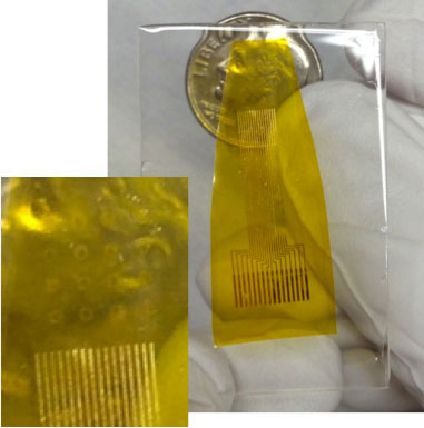
| Photograph of a 16-electrode transparent array. Inset, a closer view showing the electrode area. Fainted squares are the recording electrodes. (Image: Duygu Kuzum) | |
| Kuzum points out that, in addition to the benefits of transparency, graphene offers other advantages: It can act as an anti-corrosive for metal surfaces to eliminate all corrosive electrochemical reactions in tissues. | |
| Graphene also is inherently a low-noise material, which is important in neural recording because we try to get a high signal-to-noise ratio. | |
| To fabricate their tool, the team developed microfabrication techniques to build transparent graphene microelectrodes on flexible substrates. They transferred graphene – grown on copper by chemical vapor deposition – onto polyimide substrate with previously patterned gold contacts. Following the patterning of graphene, SU-8 was deposited as an encapsulation layer, covering the whole surface except electrode sites. | |
| "In order to investigate the feasibility of imaging through transparent doped-graphene electrodes, we performed calcium imaging in hippocampal tissue slices by both confocal and two-photon microscopy, while also conducting electrophysiological recordings," says Kuzum. "On an individual cell level, we were able to observe temporal details of seizures and seizure-like activity with very high resolution." | |
 |
|
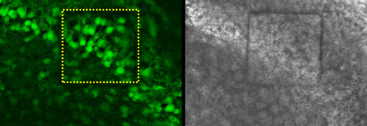
| Calcium imaging of neurons in a rat hippocampal slice through transparent graphene electrode. The square at the center, shown with yellow dashed line, is transparent graphene electrode and neurons are shown in green. (Image: Duygu Kuzum, Hajime Takano) | |
| The researchers note that the techniques developed for single electrodes in this work can easily be extended to dense arrays in order to study larger areas. | |
| "A combined sensing and imaging approach capable of simultaneously resolving both synaptic potentials and action potential firing with high spatial and temporal resolution in large populations of neurons could reveal circuit level behavior and correlation between the activities in different parts of brain," says Kuzum. "Transparent graphene electrodes are also attractive for use with optogenetics, enabling simultaneous optical stimulation of neural tissue." | |
| As the technology is further developed and used, the team expects to gain greater insight into how the physiology of the brain can go awry. For instance, it can provide information on neural circuits, which wasn't available before, because researchers didn't have the technology to probe them. That information may include the identification of specific marker waveforms of brain electrical activity that can be mapped spatially and temporally to individual neural circuits. | |
| "We can also look at other neurological disorders and try to understand the correlation between different neural circuits using this technique," says Kuzum. | |
 By
Michael
Berger
– Michael is author of three books by the Royal Society of Chemistry:
Nano-Society: Pushing the Boundaries of Technology,
Nanotechnology: The Future is Tiny, and
Nanoengineering: The Skills and Tools Making Technology Invisible
Copyright ©
Nanowerk LLC
By
Michael
Berger
– Michael is author of three books by the Royal Society of Chemistry:
Nano-Society: Pushing the Boundaries of Technology,
Nanotechnology: The Future is Tiny, and
Nanoengineering: The Skills and Tools Making Technology Invisible
Copyright ©
Nanowerk LLC
|
|
|
Become a Spotlight guest author! Join our large and growing group of guest contributors. Have you just published a scientific paper or have other exciting developments to share with the nanotechnology community? Here is how to publish on nanowerk.com. |
|