| Apr 02, 2020 |
3D reconstructions of individual nanoparticles(Nanowerk News) What do you see in the picture below? Merely a precisely-drawn three-dimensional picture of nanoparticles? Far more than that, nanotechnologists will say, due to a new study published in the journal Science ("3D structure of individual nanocrystals in solution by electron microscopy"). |
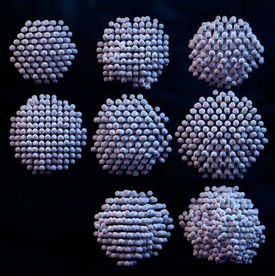 |
| Each white sphere represents the position of a platinum atom. (Image: IBS) |
| Whether a material catalyzes chemical reactions or impedes any molecular response is all about how its atoms are arranged. The ultimate goal of nanotechnology is centered around the ability to design and build materials atom by atom, thus allowing scientists to control their properties in any given scenario. |
| However, atomic imaging techniques have not been sufficient to determine the precise three-dimensional atomic arrangements of materials in liquid solution, which would tell scientists how materials behave in everyday life, such as in water or blood plasma. |
| Researchers at the Center for Nanoparticle Research within the Institute for Basic Science (IBS, South Korea), in collaboration with Dr. Hans Elmlund at Monash University's Biomedicine Discovery Institute in Australia and Dr. Peter Ercius at Lawrence Berkeley National Laboratory's Molecular Foundry in the USA, have reported a new analytic methodology that can resolve the 3D structure of individual nanoparticles with atomic-level resolution. |
| The 3D atomic positions of individual nanoparticles can be extracted with a precision of 0.02 nm--six times smaller than the smallest atom: hydrogen. In other words, this high-resolution method detects individual atoms and how they are arranged within a nanoparticle. |
| The researchers call their development 3D SINGLE (Structure Identification of Nanoparticles by Graphene Liquid cell Electron microscopy) and utilize mathematical algorithms to derive 3D structures from a set of 2D imaging data acquired by one of the most powerful microscopes on Earth. |
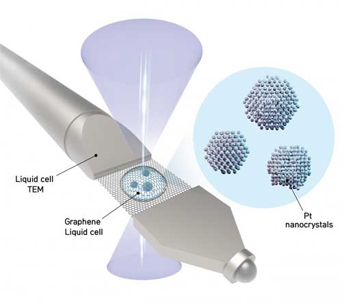 |
| The schematic shows a liquid sample contained between two sheets of graphene -- the thinnest, strongest material known. Nanoparticles in the liquid freely rotate while a transmission electron microscope takes thousands of images of the nanoparticles. The images are then analyzed by the authors' software to determine the location of every atom in each nanoparticle. (Image: IBS) |
| First, a nanocrystal solution is sandwiched in-between two graphene sheets which are each just a single atom thick. "If a fish bowl were made of a thick material, it would be hard to see through it. Since graphene is the thinnest and strongest material in the world, we created graphene pockets that allow the electron beam of the microscope to shine through the material while simultaneously sealing the liquid sample," explains PARK Jungwon, one of the corresponding authors of the study (assistant professor at the School of Chemical and Biological Engineering in Seoul National University). |
| The researchers obtain movies at 400 images per second of each nanoparticle freely rotating in liquid using a high-resolution transmission electron microscope (TEM). The team then applies their reconstruction methodology to combine the 2D images into a 3D map showing the atomic arrangement. Locating the precise position of each atom tells researchers how the nanoparticle was created and how it will interact in chemical reactions. |
| The study defined the atomic structures of eight platinum nanoparticles - platinum is the most valuable of the precious metals, used in a number of applications such as catalytic materials for energy storage in fuel cells and petroleum refinement. Even though all of the particles were synthesized in the same batch, they displayed important differences in their atomic structures which affect their performance. |
| "Now it is possible to experimentally determine the precise 3D structures of nanomaterials that had only been theoretically speculated. The methodology we developed will contribute to fields where nanomaterials are used, such as fuel cells, hydrogen vehicles, and petrochemical synthesis," says Dr. KIM Byung Hyo, the first author of the study. |
| Notably, this methodology can measure the atomic displacement and strain on the surface atoms of individual nanoparticles. The strain analysis from the 3D reconstruction facilitates characterization of the active sites of nanocatalysts at the atomic scale, which will enable structure-based design to improve the catalytic activities. The methodology can also contribute more generally to the enhancement of nanomaterials' performance. |
| "We have developed a groundbreaking methodology for determining the structures that govern the physical and chemical properties of nanoparticles at the atomic level in their native environment. The methodology will provide important clues in the synthesis of nanomaterials. The algorithm we introduced is related to new drug development through structure analysis of proteins and big data analysis, so we are expecting further application to new convergence research," notes Director HYEON Taeghwan of the IBS Center for Nanoparticle Research. |
| Source: Institute for Basic Science |
|
Subscribe to a free copy of one of our daily Nanowerk Newsletter Email Digests with a compilation of all of the day's news. |

