Showing Spotlights 457 - 464 of 559 in category All (newest first):
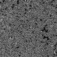 Nature has excelled in designing molecular motors, something nanotechnology researchers are still having a hard time with. The potential for nano-actuators (a nanoscale device that creates automatic motion by converting various forms of energy to rotary or linear mechanical energy) is huge - basically any active system that performs some kind of work requires an energy source. Applications reach from simple pumps on lab-on-a-chip devices to move nanoliters of fluid around to nanoscale motors for nanorobotic systems. One of the challenges of designing such a motor for the nano realm is that during the design of a nano-actuator the tradeoffs among range of motion, force, speed (actuation frequency), power consumption, control accuracy, system reliability, robustness, load capacity, etc. must be taken into consideration. Most microscale systems are currently achieved by relatively large external actuators such as syringe pumps, or high voltage power supplies, which negates the advantages of the microfabricated systems. That's why scientists are quite intrigued by the opportunity to use biological organisms to construct mechanical actuators in engineered systems at the micro- or even nanoscale. An extremely powerful biological motor is the bacterial flagellar motor found in organisms such as Escherichia coli or Serratia marcescens. Bacteria draw chemical energy directly from their environment and are able to survive in a wide range of temperature and pH. What makes bacterial propulsion system interesting for nanotechnology researchers is that bacteria are exquisitely sensitive to a wide variety of external stimuli. So far, scientists have managed to control them en masse through light (phototaxis) and chemical (chemotaxis) sensory mechanisms. In a recent example of successful use of live bacteria as mechanical actuators, scientists have built a microfluidic pump powered by self-organizing bacteria.
Jan 23rd, 2008
 Biosensors, which incorporate biological probes coupled to a transducer, have been developed during the last two decades for environmental, industrial, and biomedical diagnostics. Typically, signal sizes generated by biomolecular binding tend to be extremely small - this is the limiting factor in reaching high sensitivity for these sensors. The application of nanotechnology to biosensor design and fabrication and therapy at the molecular and cellular level promises to revolutionize bio-diagnostics. By exploiting the large surface-to-volume ratio of nanowires, nanotubes, nanocrystals, nanocantilevers, or quantum dots, researchers were able to build sensors that can measure extremely faint, and otherwise undetectable, signals - for instance a change in electrical conductance - arising from biomolecular binding on the surface of these nanodevices. Highly sensitive nanoprobes and nanosensors have the potential for a wide variety of medical uses at the cellular level. For instance, the potential for monitoring in vivo biological processes within single living cells, e.g. the capacity to sense individual chemical species in specific locations within a cell, will greatly improve our understanding of cellular function, thereby revolutionizing cell biology. One way of enhancing signal strength is on the sensor device itself and researchers have now demonstrated such an on-chip signal amplification using a standard protein on a nanoscale field effect transistor.
Jan 22nd, 2008
 For centuries, man has searched for miracle cures to end suffering caused by disease and injury. Many researchers believe nanotechnology applications in medicine may be mankind's first 'giant step' toward this goal. According to Freitas nanomedicine is "...(1) the comprehensive monitoring, control, construction, repair, defense, and improvement of all human biological systems, working from the molecular level, using engineered nanodevices and nanostructures; (2) the science and technology of diagnosing, treating, and preventing disease and traumatic injury, of relieving pain, and of preserving and improving human health, using molecular tools and molecular knowledge of the human body; (3) the employment of molecular machine systems to address medical problems, using molecular knowledge to maintain and improve human health at the molecular scale." Nanomedicine not only has the potential to change medical science dramatically but to open a new field of human enhancements that is poised to add a profound and complex set of ethical questions for health care professionals. For instance, there is a fine line between medical and non-medical uses of nanotechnology for diagnostic, therapeutic and preventive purposes (e.g. non-medical implants in soldiers). The question of whether nanotechnology should be used to make intentional changes in or to the body when the change is not medically necessary is just one hot topic in a long list of concerns. The good news is that these questions are being asked, but there is still much work to be done, but despite the enormous promise of nanomedicine, and the considerable funding going into the field, the research into the ethical, legal and social implications of nanomedicine is comparatively minute. As Peter Singer wrote in his 2003 tutorial Mind the gap: science and ethics in Nanotechnology: 'The science leaps ahead, the ethics lags behind.' As with nanotechnology in general, there is danger of derailing nanomedicine if the study of ethical, legal and social implications does not catch up with scientific developments.
Jan 8th, 2008
 1,300 to 1,400 grams and several thousand kilometers of about 100 billion interconnected nerve cells control every movement, thought, sensation, and emotion that comprise the human experience. Within the brain and spinal cord there are ten thousand distinct varieties of neurons, trillions of supportive cells, a few more trillion synaptic connections, a hundred known chemical regulating agents, kilometers of minuscule blood vessels, and untold mysteries of how - almost flawlessly - all these components work together. This is the amazing brain. Given the incredible complexity of the brain, it doesn't come as a big surprise that a lot of things can go wrong. The A-Z of brain disorders is a very long list. Several of these disorders (such as Parkinson and Alzheimer disease, but also schizophrenia, epilepsy, and bipolar disorder), not to mention tumors, are so severe that they require treatment of the brain. But even when there are promising pharmaceutical compounds for their treatment, more than 98% of these potential agents do not reach the drug development stage. The reason is the blood-brain barrier (BBB), a tight seal of endothelial cells lines the blood vessels in the brain and acts as a barrier to protect its cells. BBB strictly limits transport into the brain through both physical (tight junctions) and metabolic (enzymes) barriers and keeps most substances, such as chemicals and large biomolecules, out of the brain. The combined use of peptides and nanotechnology offers tremendous hope in the treatment of brain disorders by offering a way for drugs (the therapeutic kind) across the BBB.
Jan 7th, 2008
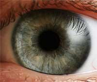 The human eye is an amazing organ. It is small, the eyeball itself weighs only about 7 grams, and it is amazingly sensitive. The eye can detect a single photon. The eye can be quicker then a race car - the young human eye can focus from infinity to 7cm in 350 milliseconds - but slow enough to witness a snail crawling across a beach. The eye can capture objects at various different angles, such as birds flying overhead or a person walking right beside you. Because the eye is such a complex optical system, it is not surprising that the list of diseases and infections that can endanger our vision is a long one. One common age-related condition is cataract. Cataract is caused by alterations in the protein structure of the lens which result in light scattering. The lens can then no longer transmit a clear picture to the retina where it can be processed and sent through the optic nerve to the brain. By age 65, over 40% of people have a cataract. Cataract is the most common cause of blindness in the world, although it is treatable. While cataract surgery is the most successful medical procedure, the inability to control penetration of the pharmacological agents into the lens and target specific intracellular biochemical pathways has impeded the success of pharmacological treatment of cataracts. Researchers are now studying the application of nanotechnology to eye lens diseases, in particular for new methods for visualizing and targeting specific intracellular mechanisms within the eye.
Jan 4th, 2008
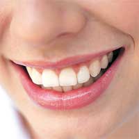 The concept of nanodentistry was introduced by Freitas in 2000: "Nanodentistry will make possible the maintenance of comprehensive oral health by employing nanomaterials, biotechnology including tissue engineering, and, ultimately, dental nanorobotics." Dental nanorobots are certainly many years off but researcher are making progress already with nanotechnology in dental care applications such as composites, bonding agents, and impression materials as well as nanostructured implant materials. For those of you with hypersensitive teeth, a new nanotechnology treatment proposed by researchers in Taiwan might one day bring pain relief. Dentine hypersensitivity (dentin is the main tissue that forms the shape of the tooth; this material exists between the pulp and the enamel, and is comprised of a series of dentinal tubules stacked on top of each other) leads to pain when fluid movement in dentinal tubules (microscopic canals that run from the outside of the dentin to the nerve inside the tooth) promotes mechanical deformation of nerve endings at the pulp/dentine interface, which is transmitted as a painful sensation. Researchers have found that sensitive teeth have an increased number of dentinal tubules (35.6% compared to 9.3%) and are wider in diameter than the dentinal tubules of non sensitive dentine. The Chinese researchers have demonstrated that this tubules can be blocked with the aid of gold nanoparticles. The world's smallest gold fillings, so to speak.
Jan 3rd, 2008
 In 1954, Richard Buckminster Fuller was granted U.S. Pat. No. 2,682,235 for geodesic domes, a method of enclosing space in architectural applications. The geodesic dome combines the structural advantages of the sphere (which encloses the most space within the least surface, and is strongest against internal pressure) with those of the tetrahedron (which encloses least space with most surface and has the greatest stiffness against external pressure). Subsequently, soccer ball shaped carbon molecules known as fullerenes or buckyballs were named for their resemblance to a geodesic sphere. But is not only certain carbon molecules where Nature uses sphere-like forms. Spheres can be found at all scales in both the inanimate and living world for the basic physical property of encapsulation. Spherical virus capsids (a capsid is the protein shell of a virus), for example, enclose space by using the geometry of the icosahedron, thus exploiting the economy of this form in terms of both surface area-to-volume ratio and genetic efficiency of subunit-based symmetric assembly. Many viruses' capsids use icosahedral symmetry to form particles ranging from 20 to 200 nanometers in size. Researchers have now begun to copy Nature's icosahedral-symmetry design principles for molecular containers, which could solve the problem of designing and synthesizing stable molecular containers having very large interior.
Dec 20th, 2007
 Past studies of photo-responsive proteins have generally dwelled on applications for energy-saving computer displays, light-based computing or computer memory. The general theme in the research tends to rely on the controllable and quick state change of the protein. But, the ability of the protein to actually incite a physical change in a composite macroscopic system has been largely unexplored. Harnessing this ability of the protein to incite change in a polymeric material could have broad implications in the field of biomedical and materials engineering. Getting bacteriorhodopsin to 'play ball' with other bulk materials could render those materials sensitive to a light. As a stimulus, light is often much easier to control than those typically explored by researchers such as bulk pH, temperature or electric fields.
Dec 13th, 2007
 Subscribe to our Nanotechnology Spotlight feed