| Posted: Dec 15, 2010 |
Nanotechnology endoscope for living cells |
| (Nanowerk Spotlight) With the advance of nanomedicine, bio-nanotechnology, and molecular biology, researchers require tools that allow them to work on a single cell level. These tools are required to probe individual cells, monitor their processes, and control/alter their functions through nanosurgery procedures and injection of drugs, DNA etc. – all without damaging the cells, of course. |
| In a previous Nanowerk Spotlight we wrote about carbon nanopipettes – glass capillaries lined with a carbon film along its inner surface and terminating with an exposed carbon nanopipe – that have been developed for this purpose. Although these pipettes can be fabricated with tips measuring well below 100 nm, their conical geometry makes it difficult to insert them into cells by more than 1 µm without causing any damage. This limits the ability to reach and interrogate organelles that are deep within the cell. |
| Other nanoscale probes based on carbon nanotubes (CNTs) attached to atomic force microscopes (see: "'Cooking' sharper AFM tips in a microwave oven") have been developed but these probes cannot handle fluids (only by dipping the probe tip into a chemical or solution and then injecting the tip into the cell). |
| Researchers have now developed a multifunctional endoscope-like device, using individual CNTs for prolonged intracellular probing at the single-organelle level, without any recordable disturbance to the metabolism of the cell. These endoscopes can transport attoliter volumes of fluid, record picoampere signals from cells, and can be manipulated magnetically. Furthermore, the tip deflects with submicrometer resolution, and the attachment of gold nanoparticles allows intracellular fingerprinting using surface-enhanced Raman spectroscopy (SERS). |
| "Our cellular endoscope is a multi-functions nanotool for studying a single cell and its organelles with high spatial and temporal resolution – all without causing damage," Michael Schrlau tells Nanowerk. "The endoscope can probe cells in much the same way that glass pipettes do – glass pipettes are widely used in cell physiology for fluid injection and to measure the electrical response of cells to stimuli. However, our endoscope goes beyond what is currently possible with existing technology: Its mechanically flexible carbon nanotube tip (<100 nm diameter) can be maneuvered remotely and enables the endoscope to pass through cell membrane unnoticed, penetrate deep into the cell, and remain inside the cell without causing harm for lengthy analysis. " |
| Schrlau, a research assistant professor at the A.J. Drexel Nanotechnology Institute explains that during these long cell penetration intervals, multiple functions are possible concurrently, including injecting attoliter volumes of fluid on demand through the nanotube, measuring electrical signals with the conductive properties of the nanotube, and taking a molecular fingerprint inside the cell with gold-decorated nanotubes. |
| Another important advantage of these novel cellular endoscopes is that they are based on glass pipettes that are used in almost every cell biology and physiology laboratory, enabling cellular endoscopes to be used with standard instruments such as manipulators, inject systems, and electrophysiology amplifiers, without requiring these laboratories to make a substantial investment in new equipment. |
| A multidisciplinary Drexel University team led by Professors Yury Gogotsi and Gary Friedman has reported their findings in a paper in the December 12, 2010 online edition of Nature Nanotechnology ("Multifunctional carbon nanotube cellular endoscopes"). |
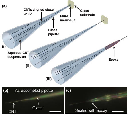 |
| Assembly and sealing of cellular endoscopes. (a) Schematic of the assembly steps: (i) pipette in contact with glass substrate surface, (ii) pipette retracted, exposing the CNT, (iii) CNT-pipette junction sealed with epoxy; (b) Optical image of a nanotube released from the template and assembled at the pipette tip. (c) Optical image of an assembled pipette sealed with epoxy resin. Scale bar in (b), (c) = 10 µm. (Reprinted with permission from Nature Publishing Group) |
| Drexel PhD student Riju Singhal fabricated the CNT-tipped endoscopes by placing multiwalled carbon nanotubes at the tip of a glass pipette using a flow-through technique. The device consisted of a carbon nanotube sealed at the end of a glass pipette tip. The epoxy-sealed carbon nanotube-glass junctions of the endoscopes were tested for robustness by repeatedly forcing them against a glass surface and then retracting them. The nanotube bent while being pushed against the glass, but recovered its shape as it was retracted |
| Schrlau notes that the endoscopes can be manufactured from nanotubes, nanowires or nanorods of any material, diameter, length or surface functionalization. |
| "Even single-walled carbon nanotubes that are capable of DNA transfer may be used as endoscope tips in some applications," he says. "In our work, we use carbon nanotubes with outer diameters ranging from 50 to 200 nm and lengths of tens of micrometers to facilitate visualization during optical microscope-based cell experiments. These nanoscopic dimensions provide sufficient mechanical strength to penetrate the membrane of the cell unnoticed, then manoeuver through the intracellular environment without inducing stress." |
| The Drexel team demonstrated several important functions of the endoscope for probing cells and they are confident that cell biologists and physiologists focusing on various fundamental biological questions will find these demonstrated capabilities of great importance. |
| Here are just a few problems the team believes can be solved with cellular endoscopes: |
| 1) Single cell analysis, such as cell response upon fluid injection and extracellular stimuli, is time-consuming and requires many data points for statistical analysis. However, the current, glass pipette-based technology often damages cells resulting in low cell survival rate and often inconclusive data from survived cells. With the minimally-invasiveness of endoscopes, the aim is to provide a more robust means of studying cells without altering normal cell behavior and increasing the throughput of analysis. Moreover, endoscopes enable analysis over longer periods of time that glass pipettes. |
| 2) The nanoscopic size of endoscopes enables them to potentially probe into some of the smaller organelles of cells, such as mitochondria and conduct multifunctional analysis: a capability elusive to the current technology. |
| 3) Endoscopes enable never-before-possible diagnostic capabilities such as gold-decorated endoscopes for intracellular surface-enhanced Raman spectroscopy (SERS), from virtually anywhere within the cell. Moreover, combinations of functions, such as injection, electrophysiology, and SERS will lead to new scientific methods and techniques as well as fundamental biological discoveries. |
| 4) In addition to biological advancements, the researchers envision components of their work will advance other fields such as micro-nanoscale device fabrication, nanochemistry and analytical methods, scanning probe microscopy and nanoscale characterization, and micro- and nanofludics. |
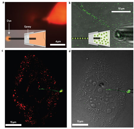 |
| Fluid and particulate flow through the nanotube endoscope. a, Optical micrographs showing the transport of an oil-based ferrofluid-fluorescent dye under an applied magnetic field. Inset: schematic of the fluid-filled nanotube endoscope ejecting fluid at its tip. b, Optical micrograph showing the flow and alignment of 50 nm polypropylene particles (green spheres) inside a carbon nanotube tip. c,d, Fluorescence (c) and differential interference contrast (d) micrographs showing a cell being interrogated with an endoscope filled with FITC-labelled (green) fluorescent polymer nanoparticles. The fluorescence micrograph in c shows mitochondria labelled with Mitotracker Orange (red). (Reprinted with permission from Nature Publishing Group) |
| Currently, the Drexel team is actively pursuing the application of cellular endoscopes for intracellular SERS and a related article has been accepted for publication and will soon appear in a premier nanobiotechnology journal. |
| "In support of our work and other carbon nanotube-based biological devices, we are taking a more detailed look at how cells respond to carbon nanotubes," says Schrlau. "Furthermore, we are currently investigating various techniques to mass produce the endoscope and transition the technology to the commercial sector. Investors are welcome!" |
 By
Michael
Berger
– Michael is author of four books by the Royal Society of Chemistry:
Nano-Society: Pushing the Boundaries of Technology (2009),
Nanotechnology: The Future is Tiny (2016),
Nanoengineering: The Skills and Tools Making Technology Invisible (2019), and
Waste not! How Nanotechnologies Can Increase Efficiencies Throughout Society (2025)
Copyright ©
Nanowerk LLC
By
Michael
Berger
– Michael is author of four books by the Royal Society of Chemistry:
Nano-Society: Pushing the Boundaries of Technology (2009),
Nanotechnology: The Future is Tiny (2016),
Nanoengineering: The Skills and Tools Making Technology Invisible (2019), and
Waste not! How Nanotechnologies Can Increase Efficiencies Throughout Society (2025)
Copyright ©
Nanowerk LLC
|

Become a Spotlight guest author! Join our large and growing group of guest contributors. Have you just published a scientific paper or have other exciting developments to share with the nanotechnology community? Here is how to publish on nanowerk.com. |
