| Posted: Mar 26, 2009 |
Tissue engineering with nanomagnets |
| (Nanowerk Spotlight) The future of tissue and cell engineering depends on the development of next-generation biomaterials that have full control over cell attachment and development into tissue. Since surface topography influences many aspects of cellular and molecular responses, surfaces of implanted devices for instance will one day be engineered to the desired cell shape and cell responses at the point of implantation. |
| The usual techniques of cell patterning are based on passive methods where the intrinsic adhesive properties of the cell are exploited. By creating substrates presenting different areas with particular adhesive characteristics (such as protein printing or 3D nanostructuring), one can segregate cells on the substrate plane. The main drawback of these techniques is their irreversibility since the differential adhesiveness is permanent. |
| Researchers in France have investigated a new direction for three-dimensional cell patterning that could find applications in tissue engineering. Rather than relying on substrate chemical or physical modifications, they perform the cell patterning using external magnetic forces with which they control the organization of cells on a substrate and create a 3D multicellular assembly. |
| "We have demonstrated an active patterning technique where the cell adhesion is guided by the application of a well-focused, local force,"Guillaume Frasca tells Nanowerk. "This force can be switched on and off at will, and when it's off the cell can behave normally, without any further modification of its metabolism." |
| Different teams have developed such strategies, mostly based on optical traps or electric forces (dielectrophoresis). The French team uses magnetic forces to guide cell adhesion. One advantage of this technique is that they can modulate the cell organization in three dimensions, while a structured substrate for passive patterning give access only to two dimensions. |
| Frasca, a PhD student at the Laboratoire Matière et Systèmes Complexes at the Université Paris-Diderot, is first author of a recent paper in Langmuir (Formation of a Three-Dimensional Multicellular Assembly Using Magnetic Patterning) that describes the 3D multicellular assembly of submillimetric dimensions, with well-defined geometry imposed by the application of a temporary constraint that does not thwart subsequent cell behavior. Frasca's PhD supervisors Florence Gazeau, and Claire Wilhelm are co-authors of the paper. The two have developed several applications in both biophysical and biomedical areas for the internalization of nanomagnets by cells. |
| "We have managed to control the organization of living cells in three dimensions with a simple protocol" says Frasca. "We only require a facile first step for magnetic labeling, and common permanent magnets to generate the magnetic forces that will drive the adhesion. Our technique is very flexible since we control a large range of experimental parameters: the intensity and geometry of the magnetic forces induced by the magnetic device; the duration of the magnetic force; the number of cells; the amplitude of the magnetic labeling; and the cell type. The magnetic field characteristics impose the geometry of the cell assembly." |
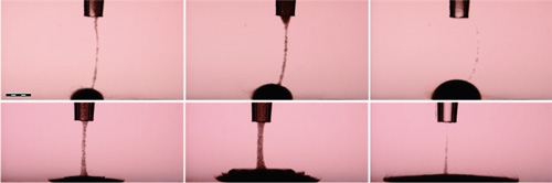 |
| Snapshots of a cell suspension (2 x 10⁵ cells in 1 µL) released from a micropipette (time interval ) 2s). (Top) Magnetically labeled cells are massively attracted by the magnetized tip and pile up to form a 3D multicellular assembly of controlled dimensions. (Bottom) By contrast, the same cell suspension spreads over the substrate if no magnetic force is applied. Bar ) 500 µm. (Reprinted with permission from American Chemical Society). |
| This technique is based on an ubiquitous, non-toxic magnetic labeling of mammalian cells by natural internalization of maghemite nanoparticles. These nanomagnets made of iron oxide – easily synthesized in the lab – are covered by an anionic shell. Frasca explains that this structure gives them the ability to develop electrostatic interactions with the plasma membrane which enable direct internalization by the cell and subsequent concentration in natural vesicles called endosomes. |
| "These nanomagnets could challenge the use of commercial nanoparticles, which are covered with a layer of polymers and thus require the use of a transfecting agent to enter cells," he says. "We have already tested this labeling method on a large number of cell types from different species, immune cells, malignant cells, muscle cells, etc (see Universal cell labeling with anionic magnetic nanoparticles for a review). The labeling did not affect cell proliferation, or the therapeutic and functional properties of the tested cell types, either in vitro or in vivo. These magnetic cellular markers are therefore fully biocompatible." |
| Research findings like this one by the French scientists are proof-of-principle studies. The main challenge for tissue engineering, however, is the need to scale these techniques from thousands of cells up to the vastly bigger dimensions of entire organs. |
| Frasca also points out that research teams need to increase the cooperation between physicians, biologists and physicists in order to gain a better understanding of the processes at stake in the differentiation of stem cells – a critical step for the formation of tissues. |
 By
Michael
Berger
– Michael is author of four books by the Royal Society of Chemistry:
Nano-Society: Pushing the Boundaries of Technology (2009),
Nanotechnology: The Future is Tiny (2016),
Nanoengineering: The Skills and Tools Making Technology Invisible (2019), and
Waste not! How Nanotechnologies Can Increase Efficiencies Throughout Society (2025)
Copyright ©
Nanowerk LLC
By
Michael
Berger
– Michael is author of four books by the Royal Society of Chemistry:
Nano-Society: Pushing the Boundaries of Technology (2009),
Nanotechnology: The Future is Tiny (2016),
Nanoengineering: The Skills and Tools Making Technology Invisible (2019), and
Waste not! How Nanotechnologies Can Increase Efficiencies Throughout Society (2025)
Copyright ©
Nanowerk LLC
|

Become a Spotlight guest author! Join our large and growing group of guest contributors. Have you just published a scientific paper or have other exciting developments to share with the nanotechnology community? Here is how to publish on nanowerk.com. |
