Nanoscopy Techniques: Breaking the Diffraction Limit in Microscopy
What is Nanoscopy?
Nanoscopy is a field of microscopy that focuses on imaging and studying structures and processes at the nanoscale, typically below the diffraction limit of light. It encompasses various techniques that overcome the limitations of conventional optical microscopy, enabling the visualization and analysis of nanoscale features with unprecedented resolution and precision.
Breaking the Diffraction Limit
One of the main challenges in conventional optical microscopy is the diffraction limit of light, which restricts the spatial resolution to about half the wavelength of the illuminating light (typically around 200-300 nm). This limitation prevents the visualization of nanoscale features that are smaller than this limit. Nanoscopy techniques aim to break this diffraction barrier and achieve super-resolution imaging.
Nanoscopy Techniques
Several nanoscopy techniques have been developed to overcome the diffraction limit and enable nanoscale imaging:
Stimulated Emission Depletion (STED) Microscopy
STED microscopy uses a doughnut-shaped depletion laser to selectively suppress the fluorescence emission from the periphery of the excitation spot, effectively reducing the size of the point spread function (PSF). By scanning this smaller PSF across the sample, STED can achieve lateral resolutions of around 20-50 nm.
Single-Molecule Localization Microscopy (SMLM)
Single-Molecule Localization Microscopy techniques, such as Photoactivated Localization Microscopy (PALM) and Stochastic Optical Reconstruction Microscopy (STORM), rely on the stochastic activation and precise localization of individual fluorescent molecules. By repeatedly activating and imaging sparse subsets of molecules, SMLM can reconstruct super-resolved images with resolutions of around 10-20 nm.
Structured Illumination Microscopy (SIM)
Structured Illumination Microscopy uses patterned illumination to encode high-frequency spatial information into the observed image. By capturing multiple images with different illumination patterns and applying computational reconstruction algorithms, SIM can double the lateral resolution compared to conventional microscopy, achieving resolutions of around 100-130 nm.
Applications of Nanoscopy
Nanoscopy has found wide-ranging applications in various fields, including:
Life Sciences
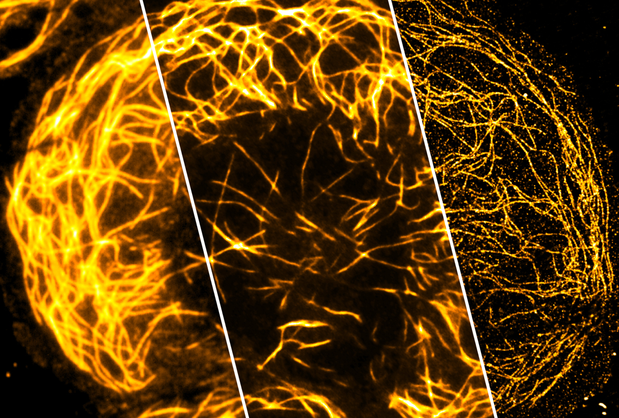
Nanoscopy has revolutionized the study of biological systems at the nanoscale. It enables the visualization of subcellular structures, protein complexes, and molecular interactions with unprecedented detail. Nanoscopy techniques have shed light on the organization and dynamics of cellular components, such as the cytoskeleton, membrane receptors, and organelles.
Materials Science
Nanoscopy is a powerful tool for characterizing nanomaterials and understanding their properties and behavior at the nanoscale. It allows researchers to study the morphology, composition, and defects of nanostructures, such as nanoparticles, nanowires, and thin films. Nanoscopy techniques can provide valuable insights into the structure-property relationships of nanomaterials and guide their design and optimization.
Nanoelectronics and Nanophotonics
Nanoscopy plays a crucial role in the development and characterization of nanoelectronic and nanophotonic devices. It enables the imaging and analysis of nanoscale components, such as transistors, waveguides, and photonic crystals, with high spatial resolution. Nanoscopy techniques can help in understanding the operation, performance, and limitations of these devices and facilitate their optimization and integration.
Challenges and Future Perspectives
Despite the remarkable advancements in nanoscopy, several challenges remain to be addressed. One of the main challenges is the trade-off between resolution, imaging speed, and sample compatibility. Achieving high resolution often requires longer acquisition times and may be limited to specific sample preparations. Improving the temporal resolution and extending nanoscopy to live-cell and in vivo imaging are active areas of research.
Future developments in nanoscopy will focus on pushing the resolution limits even further, approaching the single-molecule level. The integration of nanoscopy with other imaging modalities, such as electron microscopy and atomic force microscopy, will provide a more comprehensive understanding of nanoscale systems. Additionally, the development of advanced computational methods, such as machine learning and artificial intelligence, will enhance the image reconstruction, analysis, and interpretation capabilities of nanoscopy.
Further Reading
Current Biology, Force nanoscopy of living cells
IEEE Access, Smart Nanoscopy: A Review of Computational Approaches to Achieve Super-Resolved Optical Microscopy
Science, Far-Field Optical Nanoscopy
