| Dec 16, 2019 |
Living biosensors and micromotors as platform for precision medicine |
| (Nanowerk Spotlight) Theranostics – a combination of the words therapeutics and diagnostics – describes an integrated therapeutic platform for cancer treatment that can diagnose a tumor, deliver targeted therapy, and monitor the response to therapy in a single procedure. Theranostic medicine is a step towards personalized medicine and has the potential for simultaneous and real-time monitoring of drug delivery, trafficking of drug, and therapeutic responses. |
| Optical biosensors and micromotors that can sense and actuate biological environments are considered promising theranostic devices because of their high spatial resolution and flexibility. |
| Advancing the field from synthetic sensors to living materials – which often can be incompatible with biological systems – researchers in China have assembled living biosensors and micromotors using an in vivo red blood cell (RBC) waveguide for applications in pH sensing and particle transport. |
| The scientists reported their results in Advanced Functional Materials ("Red-Blood-Cell Waveguide as a Living Biosensor and Micromotor"). |
| "Our living biosensor can be used for the diagnosis of pH-related disorders of the blood; then, the waveguide can rotate as a micromotor and transport microparticles to the target region," Yuchao Li, an associate professor at the Institute of Nanophotonics, Jinan University, and the paper's first author, tells Nanowerk. "Compared with synthetic sensors and motors, the RBC biosensor and micromotor are highly biocompatible, flexible, and noninvasive." |
| The team confined their RBC waveguide within the optical axes of two tapered fiber probes (which could be used to trap cells, guide light, and collect signals) via an optical gradient force. The light propagation mode of the RBC waveguide was related to the RBC morphology, which depends on the pH of blood. |
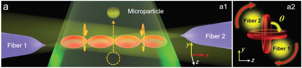 |
| Schematic diagrams show optical transport of a microparticle in a microfluidic capillary with a1) the RBC micromotor, which is driven by an optical torque induced by the coherent output light from a2) two fiber probes. Red arrows indicate the rotation direction and θ denotes the rotation angle. (Reprinted with permission from Wiley-VCH Verlag) (click on image to enlarge) |
| Therefore, the RBC waveguide could be used for pH sensing and then to reveal pH-induced diseases of the blood. After sensing, optical torque (induced by the coherent output light from the two fiber probes) was applied to the RBC waveguide, allowing it to continuously rotate to transport microparticles to a target site inside a microfluidic capillary. |
| As can be seen in the video below, during rotation of the RBC micromotor, the microparticles inside the capillary are transported forward because the RBC micromotor could induce local flow with its driving force. |
| The rotation speed of the micromotor is linearly proportional to the input optical power with a maximum speed of 560 rpm achieved at 50 mW. At 15 mW, the rotation speed is 150 rpm, which transports a microparticle 25 µm in 1.5 seconds. |
| To extend its potential applicability, the team also successfully assembled and operated the RBC waveguide in zebrafish blood vessels. |
| "Compared with in vitro assembly of the RBC waveguide, in vivo assembly is more difficult because of the complex physiological environments inside biosystems, including the blood stream and high-scattering tissues," Li points out. |
| For their experiments, the team chose zebrafish for the in vivo assembly of the RBC waveguide because their tails are optically transparent and their blood vessels can thus be observed under an optical microscope. |
| As illustrated in the figure below, two laser beams at 980 nm were focused on a blood vessel in the fish's tail by fiber probes, which were placed above the fish with the probe tips approximately 10 µm away from the surface of the skin. |
| The red blood cells in the blood vessel of the fish become trapped in the focus of the laser beam and bound into a 1D waveguide by an optical gradient force. |
 |
| Schematic diagram showing optical assembly of the RBC waveguide inside a zebrafish. The inset shows an optical image of the zebrafish used in the experiments. b) Schematic diagram showing optical assembly of the RBC waveguide inside the blood vessels. (Reprinted with permission from Wiley-VCH Verlag) |
| "Our experiment demonstrates that the RBC waveguide can be controllably assembled in a living vessel, and highlights the in vivo potential of the proposed technique," Li concludes. "After our experiments using zebrafish we will next try to assemble a biosensor and micromotor inside the human body." |
 By
Michael
Berger
– Michael is author of four books by the Royal Society of Chemistry:
Nano-Society: Pushing the Boundaries of Technology (2009),
Nanotechnology: The Future is Tiny (2016),
Nanoengineering: The Skills and Tools Making Technology Invisible (2019), and
Waste not! How Nanotechnologies Can Increase Efficiencies Throughout Society (2025)
Copyright ©
Nanowerk LLC
By
Michael
Berger
– Michael is author of four books by the Royal Society of Chemistry:
Nano-Society: Pushing the Boundaries of Technology (2009),
Nanotechnology: The Future is Tiny (2016),
Nanoengineering: The Skills and Tools Making Technology Invisible (2019), and
Waste not! How Nanotechnologies Can Increase Efficiencies Throughout Society (2025)
Copyright ©
Nanowerk LLC
|

Become a Spotlight guest author! Join our large and growing group of guest contributors. Have you just published a scientific paper or have other exciting developments to share with the nanotechnology community? Here is how to publish on nanowerk.com. |
