| Posted: Jun 30, 2016 | |
Graphene coated microbubbles as superior photoacoustic imaging contrast agent |
|
| (Nanowerk Spotlight) Researchers have demonstrated that the coupling of pristine graphene sheets on practically any polymer surface can be accomplished in mild reaction conditions and in aqueous medium. The method leaves intact the 2D planar structure of graphene preserving its original features. | |
| This novel hybrid construct enables in vivo photoacoustic signal enhancement and is a very promising step forward for an implementation of photoacoustic imaging (PAI), a powerful preclinical diagnostic tool. | |
| Imaging and drug delivery based on miniaturized devices are keys in the future of personalized medicine. One of the main issues is the disease detection in the earliest stages. This increases the chance of success of any therapy. PAI is among the imaging methods with the highest resolution and this allows a less invasive way to detect tumors at very early stages. The targeting is key both for a localized diagnostic and to bring a drug focally to the diseased tissue. | |
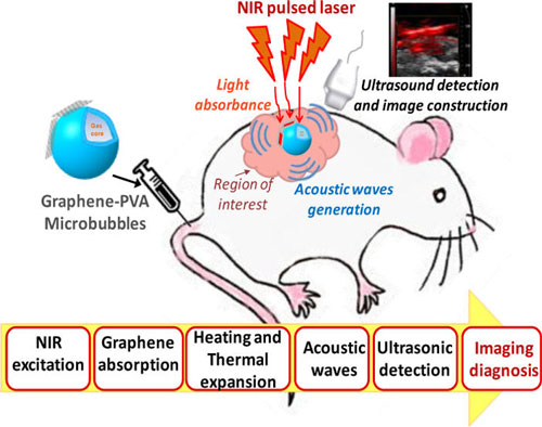
| The photoacoustic effect, discovered and studied by Bell more than 135 years ago (Nature, "Selenium and the Photophone"), occurs when light hits an absorber and the locally accumulated thermal energy is converted and dissipated in mechanical energy by the emission of ultrasound waves and detected by a transducer. | |
| The light wavelength used in biomedical diagnostics is in the near infrared (NIR) spectral window, where light is less attenuated by the tissue (and water). Endogenous metabolites such as haemoglobin of red blood cells behave in this way. | |
| "This effect is used in photoacoustic imaging (PAI) and it can be enhanced by exogenous devices as for example our hybrid assembly made by the stable coupling of pristine graphene with microbubbles," Gaio Paradossi, a professor in the Dipartimento di Scienze e Tecnologie Chimiche at Università di Roma Tor Vergata, explains to Nanowerk. "The efficiency in the enhancement of the photoacoustic signal makes such device an unprecedented multimodal contrast agent for ultrasound and PAI." | |
| Paradossi and his team just reported in ACS Applied Materials & Interfaces ("Graphene Meets Microbubbles: A Superior Contrast Agent for Photoacoustic Imaging") a proof of concept, tested in vivo, where they fabricated a hybrid injectable device for use as an efficient and versatile photoacoustic contrast agent. | |
 |
|
| Schematics of the approach. (Reprinted with permission by American Chemical Society) | |
| In their present work, the researchers present a technique to couple pristine graphene with polymer shelled microbubbles. The design is based on poly(vinyl alcohol) (PVA) polymer microbubbles, which are stably coupled to pristine graphene sheets through surfactant moieties covalently bound to the available functional groups on the microbubbles surface. | |
| "At the center of our work is pristine graphene, the intact form of graphene," Paradossi points out. "Most of the applications reported in the literature highlight the use of graphene oxide (GO) or reduced graphene oxide (rGO). These forms of graphene, not directly obtained by graphite exfoliation, derive from chemical modifications of the 2D structure of graphene in very harsh conditions, which introduce kinks and irregularities in the carbonic material." | |
| "Such modifications make GO and rGO more reactive and more processable than pristine graphene, but jeopardize the electrical, optical and mechanical properties of this material," he adds. | |
| In their present work, the researchers present a technique to couple pristine graphene with polymer shelled microbubbles. | |
| "Why are polymer shelled microbubbles such an exotic support for pristine graphene? Microbubbles are the best contrast agents for enhancing ultrasounds and it is a natural choice if ultrasound or photoacoustic imaging are the goals," says Paradossi. "Another important issue pointed out in our paper is the exceptional stability of the coupling to the polymer surface of the microbubbles is an asset for the biocompatibility of graphene." | |
| This work contains several novel elements: | |
|
|
|
| These results have been a by-product of the work presently carried out within the frame of the European project TheraGlio – Developing theranostics for Gliomas, where the goal is to develop a multimodal imaging system for Theranostics (therapy + diagnosis) of patients bearing malignant glioma, the most common primary brain tumour. | |
 |
|
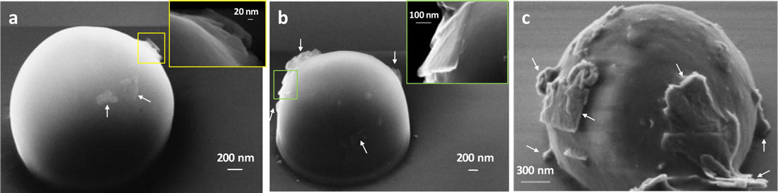
| FESEM images of (a) G/PVA 2.5% (w/w), (b) G/PVA 5% (w/w), and (c) G/PVA 10% (w/w); insets, magnified graphene flakes on PVA microbubble shell of the selected zones. The arrows indicate graphene sheets. (Reprinted with permission by American Chemical Society) (click on image to enlarge) | |
| The results also build on a method recently developed by Paradossi's group where graphene sheets were stably anchored to PVA hydrogels (The Journal of Physical Chemistry B, "Soft Confinement of Graphene in Hydrogel Matrixes"). This method consists of the ultrasound exfoliation of graphite assisted by a surfactant in aqueous medium followed by the tethering to the polyvinyl alcohol chemical hydrogels via the surfactant functional moieties. | |
| Going forward, the team will address the biocompatibility of their graphene microbubbles; the ability to target pathological cells tissues; and ultrasound assisted drug delivery. | |
| As for biocompatibility, graphene is anchored to the surface of the PVA shelled microbubble in a stable way and loss of graphenic material was not monitored in physiological media. PVA is already known as a biocompatible polymer and it has been used for the fabrication of echogenic microbubbles with long shelf-life and good chemical versatility (see: Gaio Paradossi “Hydrogels Formed by Cross-linked Poly(vinyl alcohol)” in Polymeric Biomaterials: Structure and Function, Volume 1). | |
| "However, for such hybrid system an increase of biocompatibility should be expected by surface pegylation," says Paradossi. "The chemical versatility of the shell can allow tumor tissues to be targeted by conjugating the peptide sequences as cyclic RGD or hyaluronic acid on the PVA microbubble surface. RGD is known to bind the receptor of αVβ3 integrins, a family of membrane proteins, which is over expressed by tumor cells." | |
| "Analogously, grafting hyaluronic acid, a polysaccharide present in the extracellular matrix of mammals, on the PVA surface is a mean to address the graphene/microbubble device on the receptor of CD44, another membrane protein over expressed by tumor cells." | |
| The microbubbles can also be converted to drug delivery systems by loading drugs directly on the surface by physisorption. Ultrasound can be used to excite the microbubbles – to 'shake' them – and release the drug molecules. | |
| "More sophisticated methods are under study in our lab, consisting in tethering liposomes on the shell containing oligonucleotides cargo which can be transfected upon ultrasound irradiation," Paradossi notes. | |
| In conclusion, anchoring graphene on PVA microbubble surfaces opens the way to leap from the use in small size animals functional imaging to a high resolution clinical diagnostic tool, by combining the appealing features of both PVA microbubble (as efficient ultrasound scatterer) and graphene (as strong NIR absorber with high thermal conductivity). | |
 By
Michael
Berger
– Michael is author of three books by the Royal Society of Chemistry:
Nano-Society: Pushing the Boundaries of Technology,
Nanotechnology: The Future is Tiny, and
Nanoengineering: The Skills and Tools Making Technology Invisible
Copyright ©
Nanowerk LLC
By
Michael
Berger
– Michael is author of three books by the Royal Society of Chemistry:
Nano-Society: Pushing the Boundaries of Technology,
Nanotechnology: The Future is Tiny, and
Nanoengineering: The Skills and Tools Making Technology Invisible
Copyright ©
Nanowerk LLC
|
|
|
Become a Spotlight guest author! Join our large and growing group of guest contributors. Have you just published a scientific paper or have other exciting developments to share with the nanotechnology community? Here is how to publish on nanowerk.com. |
|