Showing Spotlights 97 - 104 of 110 in category All (newest first):
 It has been 25 years since the scanning tunneling microscope (STM) was invented, followed four years later by the atomic force microscope, and that's when nanoscience and nanotechnology really started to take off. Various forms of scanning probe microscopes based on these discoveries are essential for many areas of today's research. Scanning probe techniques have become the workhorse of nanoscience and nanotechnology research. Given the 25-year development timeframe, it is surprising that even today there is no generally accepted standard for scanning probe microscopy (SPM). There is no unified SPM terminology, nor is there a standard for data management and treatment, making access and processing of SPM data collected by different types of instruments an error-prone exercise. SPM standardization has only recently begun as part of an effort by the International Organization for Standardization (ISO) the largest developer of industrial standards. Meanwhile the development of SPM instruments and analysis software continues, increasing the already large family of scanning probe microscopy.
May 25th, 2007
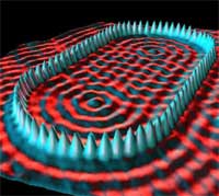
 Magnetic resonance imaging (MRI) is a powerful imaging technology that serves as a non-invasive method to render images of the inside of an object. It is primarily used in medical imaging to demonstrate pathological or other physiological alterations of living tissues. MRI also has uses outside of the medical field, for instance as a non-destructive testing method to characterize the quality of products such as produce and timber. Conventional MRI usually operates at the scale of millimeters to micrometers - 3 micrometers at best - which is good enough for the mostly medical diagnostic purposes it is used for. Researchers have now shown that the imaging of nuclear spins using magnetic resonance, the basis for MRI, can be pushed to sub-100nm resolution into the nanoscale realm. They demonstrated that using an emerging technique based on force detection, they can image nuclear spins with a sensitivity that is 60,000 times better than MRI. The resolution is about 30 times better than the most advanced conventional MRI imaging. By improving this technique, researchers will be able to push deeper into the nanometer regime and approach the capability needed for direct three-dimensional imaging of individual macromolecules.
May 3rd, 2007
 It its more than 25 years of existence, Scanning Tunneling Microscopy (STM) has predominantly brought us extremely detailed images of matter at the molecular and atomic level. STM - not to be confused with the scanning electron microscope (SEM) - is a non-optical microscope that scans an electrical probe over a surface to be imaged to detect a weak electric current flowing between the tip and the surface. The STM allows scientists to visualize regions of high electron density and hence infer the position of individual atoms and molecules on the surface of a lattice. Researchers have now taken a further step by using STM to perform real-time single-molecule imaging of an entire chemical reaction. Many chemical reactions are catalyzed by metal complexes, and insight into their mechanisms is essential for the design of future catalysts. These new findings have demonstrated that the STM approach to studying chemical reactions in a dynamic environment can provide valuable information about reaction mechanisms and rates, as well as catalyst activity and stability.
May 2nd, 2007
 Synthesis of carbon nanotubes (CNTs) is a rapidly advancing field, but there is a lot that researchers don't know about how nanotubes form and grow. Synthesis, while rapidly developing, is currently the weakest link for most nanotube applications, with high yield and high precision diameter and chirality control being important goals. Historically, in situ characterization tools have accelerated progress in synthesis for many advanced materials, and there is widespread recognition that in situ tools have the potential to improve CNT synthesis as well. Ideally one would like to detect individual nanotubes and ensembles as they grow and measure their physical properties while imposing minimal constraints on the synthesis method. In other words, with a good understanding of the synthesis process we would be better able to control the product. It is feasible that by actually observing nanotubes as they grow one will gain a better understanding of the growth process and also better characterize the grown product. Greater control over the physical characteristics of the nanotube product is essential to enable many applications, as well as many fundamental studies. Although chemical vapor deposition (CVD) is now a very standard method to synthesize CNTs, there aren't really standard in situ tools to characterize nanotubes during growth. Researchers in Canada have now shown how global Raman imaging (GRI) can be used to characterize the CVD growth of CNTs in situ and in real time.
Apr 26th, 2007
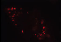 Fluorescent semiconductor nanocrystals (or quantum dots) hold a number of advantageous features including high photobleaching thresholds and broad excitation but narrow emission spectra well suited for multicolor labeling and detection. Unfortunately, most quantum dots are toxic, and hence reduction of cytotoxicity and human toxicity through surface modification plays a pivotal role in their successful application to in vivo labeling, imaging, and diagnosis. Researchers now have demonstrated that nanodiamond particles possess several unique features, including facile surface modification, long-term photostability, and no fluorescence blinking, that makes their detection and long-term tracking in living cells not only possible but practical.
Mar 1st, 2007
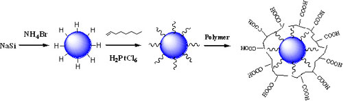 Current medical and biological fluorescent imaging is limited by the use of dye markers, which are not photostable. The dyes can break down under photoexcitation, room light or higher temperatures. The observation of strong visible emission in porous silicon therefore has triggered substantial interest in exploring the synthesis and characterization of silicon nanoparticles. Due to their biocompatibility, high photoluminescence quantum efficiency and stability against photobleaching, silicon nanoparticles are expected to be an ideal candidate for replacing fluorescent dyes in many biological assays and fluorescence imaging techniques. For instance, they have been proposed as better quantum dots for in vivo applications, potentially replacing quantum dots of highly toxic cadmium. Different synthetic and physical methods have been used to prepare silicon nanoparticles. However, the yields of nanoparticles from these methods are very low and an HF (hydrofluoric acid) etching process is often necessary to obtain photoluminescent, hydrogen-terminated silicon nanoparticles. Now, researchers have developed a new solution route for the production of macroscopic amounts of hydrogen terminated silicon nanoparticles without hazardous material handling. This synthesis route is simple and thus offers great opportunity for scaled-up preparation of semiconductor materials.
Feb 19th, 2007
 In recent years, the manipulation of single atoms and molecules has been a major advance in the application of the scanning tunneling microscope (STM). The main appeal of STM manipulation is the ability to access, control and modify the interactions between the tip and the adsorbate, a few angstroms apart. So far, however, atom manipulation using a STM or an AFM -tip has been restricted to flat surfaces. Manipulation of atoms on a rough terrain requires much more precise control at the atomic scale. Researchers now report extraction and manipulation of individual silver atoms on three dimensional silver nanoclusters. This is the first demonstration that individual atoms can be repeatedly pulled out from a silver cluster on a silver surface using STM tip. It is also the first atom manipulation work done on a 3-D surface. There are still very few research groups that have demonstrated single atom manipulation with atomic scale precision on flat surfaces. This remarkable achievement has an impact on the fundamental understanding of interactions between the matters. While it certainly is not a commercial production technique, it does further the fundamental understanding of the interaction between atoms, and it is an atom production technique that can be used to extract the atoms for atomistic construction.
Dec 19th, 2006
Conventional diagnostic imaging is mainly based on morphological contrast that is a result of different general tissue characteristics. Molecular imaging is a new approach for detecting diseases much earlier, visualizing biological processes at the cellular and molecular level in living organisms, and detecting changes in biochemistry. Corresponding molecular markers appear in quite low concentrations. Hence, the imaging technique must be very sensitive. Magnetic resonance imaging (MRI) has some significant advantages in terms of using non-ionizing radiation (in contrast to x-rays) and giving high resolution tomographies for any arbitrary position and orientation. However, conventional MRI suffers from inherent low sensitivity. A new method, using xenon as the signal source, was developed by researchers in California and will make MRI an important technique in molecular imaging, offering a huge potential for specific detection of disease markers. The new technique allows detection of signals from molecules present at 10,000 times lower concentrations than conventional MRI techniques. Called HYPER-CEST, for hyperpolarized xenon chemical exchange saturation transfer, this new technique could become a valuable tool for medical diagnosis, including the early detection of cancer.
Nov 7th, 2006
 Subscribe to our Nanotechnology Spotlight feed