Showing Spotlights 1 - 8 of 110 in category All (newest first):
 New microchip design with 10-nanometer windows facilitates unprecedented electron microscope imagery resolution to directly visualize chemical reactions at fundamental scales.
Jan 22nd, 2024
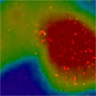
 New microscopy techniques now enable direct imaging of atomic defects' electric field polarization, unlocking understanding of defect tuning mechanisms for enhanced electrocatalysis and beyond.
Dec 17th, 2023
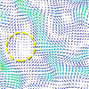
 Super-resolution microscopy has transformed the way we view the biological world, and enabled scientists to visualize biological structures and their interactions inside cells, at a resolution of a few nanometers. But 'seeing' atomic motion in space and time is not straightforward. Researchers set out do something similar and see how individual atoms move in a molecule using X-rays. The result is an approach to extend super-resolution imaging to the challenging case of ultrafast scattering.
Mar 8th, 2023
 Every cell relies on the uptake (endocytosis) of materials like proteins, cytokines and even synthetic carbon nanomaterials, to perform its required cellular fate functions. Studying this process in detail is an extremely challenging and thus extremely interesting goal in biophysics. Therefore, endocytosis is of interest for bringing therapeutic targets into cells. Studying the pathways of how materials get into the cell can aid in untangling trafficking to design higher efficiency targeted drug and gene delivery therapies.
Jun 20th, 2022
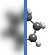
 Scanning probe lithography techniques rely on the use of cantilevers to pattern sub-100 nm structures generated by the mechanical contact between a cantilever tip and a surface. SPL, with its high resolution, is a popular method for prototyping nanoscale structures. Overcoming the current limitations of SPL - the lack of high-resolution, high-speed throughput at low cost - researchers developed a new technique they termed nanocalligraphy scanning probe lithography.
Oct 28th, 2021
 Relying on the quantum confinement effect, the strong light-matter interaction in low-dimensional materials enables them to exhibit excellent photodetection. The unique out-of-plane van der Waals force in low-dimensional layered materials makes them free from the surface dangling bonds compared to traditional bulk materials, which reduces the dark current of the devices by eliminating surface recombination. These unique advantages make low-dimensional materials have the potential to achieve breakthroughs in the field of low-cost high-performance room-temperature infrared detection.
Jun 7th, 2021
 Taking advantage of its piconewton force and sub-nanometer displacement resolution, atomic force microscopy (AFM) is uniquely suited to measure nanoscale mechanical properties, especially when it comes to soft materials. Force spectroscopy is a useful nanomechanical technique to obtain both single point measurements and maps of important mechanical properties such as stiffness and adhesion. Cantilever and tip calibrations coupled with contact mechanics models enable the full analysis and interpretation of individual force curves.
Oct 30th, 2020
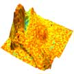
 High-entropy alloys (HEAs), which are formed by combining nearly equal parts of several primary metals, are an emerging class of advanced materials that hold great potential for creating materials with superior mechanical, thermal, and catalytic properties. New research offers key insights into how HEA nanoparticles behave under high-temperature oxidizing environment and sheds light on future design options of highly stable alloys under complex service conditions.
Oct 29th, 2020
 Subscribe to our Nanotechnology Spotlight feed