Showing Spotlights 33 - 40 of 110 in category All (newest first):
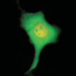
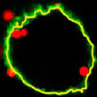 While nanoparticles are emerging as drug carriers for targeted nanomedicines, preclinical assays to test nanoparticle efficacy are hampered by the lack of methods to quantitatively determine internalized particles. A novel method is suited to pave the way for preclinical testing of nanoparticles to establish dose-efficacy relationships and to optimize biophysical and biochemical parameters in order to make better drug delivery vehicles. The team demonstrated that it is possible to determine the exact number of nanoparticles inside a cell through a combination of three methods and a mathematical model which they developed to link the data from these three methods.
Jun 18th, 2013
 Ferromagnetic materials exhibit the so-called anomalous Hall effect (AHE), whereby the electrons flowing through the material experience a lateral force pushing them to one side as a result of the material's intrinsic magnetization. Although the AHE has been used in the field on nanotechnology to measure the magnetic behavior of nanoparticles (with sizes larger than 50 nm), nobody so far had tried to separate the signals of the individual particles. Researchers in Germany have now developed a simple technique which allows to measure the magnetic response of single ferromagnetic nanoparticles down to a radius of about 3.3 nm.
May 14th, 2013
 Nanopores are an exciting class of single molecule nanosensors. For several years now, nanopore technology has been developed as a biosensor at the single-molecule resolution to detect an array of biomedical molecules, such as DNA, RNA, protein, biotoxin, and various nanopore projects have been funded to develop the next generation of DNA sequencing technology. The sensing principle is based on the resistive pulse technique - molecules are detected as they pass through a single nanopore since during translocation the molecules exclude ions and therefore modulate the current. In new work, researchers have demonstrated the single molecule detection of a wide range of proteins with solid state glass nanopores.
May 9th, 2013
 For a transistor to work properly, it must contain impurity atoms - called dopants - replacing the silicon atoms at certain places in the device. Given that modern transistor are approaching the atomic scale, the exact location of a single dopant atom becomes critical in determining the device functionality. In a different context, single dopant atoms in semiconductors have now proved to be an excellent platform to encode quantum information. Therefore, the exact location of single dopant atoms is also crucial to future quantum computers based on silicon. A new technique allows the accurate location of a single dopant atom in a nanoscale device, after the device has been fabricated, and without damaging or altering any of its functionalities.
May 8th, 2013
 Most of the research efforts on developing synthesis methods for graphene has focused on flat substrates. However, direct growth of graphene layers on prepatterned substrates has remained elusive. In new work, resarchers have grown graphene in prepatterned copper-coated substrates, and they apply this protocol for the fabrication of MEMS devices, in particular, atomic force microscope probes. This layer of graphene improves the functionality of the probes by making them conductive and more resistant to wear.
May 6th, 2013
 Applications and studies in DNA-based biophysics, biochemistry and biotechnology rely on accurate imaging with high temporal and spatial resolution towards the mesoscopic and single-molecule levels. This, in turn, relies on the ability to immobilize and stretch portions of DNA on a substrate without damaging it. Researchers in Italy have now reported a noninvasive, all-optical, holographic technique for permanently aligning liquid crystalline DNA filaments in a microperiodic template realized in soft-composite materials.
Mar 25th, 2013
 Researchers are very interested in investigating the biomechanical properties of the inner structure of cells due to their relevance in many important topics in biology such as intracellular and intercellular dynamics; tissue and organs formation and their homeostasis; but also in medicine as the formation and development of diseases like inflammatory disorders or tumor. In order to study inner cell properties, researchers have now presented a biophotonic holographic workstation that combines the complementary features of holographic optical tweezers (HOT) and self-interference digital holographic microscopy, in order to investigate biomechanics properties at the single cell level.
Dec 7th, 2012
 Fluidic force microscopy (FluidFM) is an emerging technology which combines atomic force microscopy (AFM) with microfluidics. In a new study, researchers in Switzerland have now developed an innovative method for straightforward injection into the nucleus of a living cell, taking advantage of the nanoscale accuracy and small probe size of AFM and the possibility to handle fluid under pressure-control through the integrated microchannel.
Nov 29th, 2012
 Subscribe to our Nanotechnology Spotlight feed