| Mar 16, 2026 |
Silicon nanotube arrays deliver mRNA into human stem cells while preserving pluripotencySilicon nanotube arrays deliver functional mRNA into human pluripotent stem cells for the first time, achieving up to 64% transfection without compromising stem cell identity. |
| (Nanowerk Spotlight) Gene therapies, cell-based treatments, and gene-editing tools like CRISPR all depend on one thing: getting nucleic acids into living cells reliably and safely. Viral carriers do this efficiently but can integrate into the genome, raising long-term safety concerns. Chemical and electrical methods avoid that risk but often damage cells or work inconsistently. |
| A physical alternative now exists. Arrays of nanoscale hollow needles, fabricated from silicon or polymers, allow cells to be pressed onto structures that penetrate the membrane and release molecular cargo directly into the cytoplasm. This approach, known as nanoinjection, has already proven effective for engineering immune cells, fibroblasts, and neurons with minimal toxicity. It does not, however, work on human induced pluripotent stem cells (hiPSCs), the very cells that stand to benefit most. |
| These reprogrammed adult cells can become virtually any cell type in the body, making them indispensable for disease modeling, drug discovery, and regenerative medicine. But they are also among the most temperamental cells in any laboratory. They require extracellular matrix coatings to adhere, colony-based growth to survive, and exact handling conditions at every step. Minor disruptions trigger cell death or, worse, quietly erase the pluripotency that gives them their value. |
| A study now published in Advanced Materials ("Poking Pluripotency: Nanoinjection Into Human iPSCs") reports how a team based primarily at Monash University and Deakin University in Australia overcame this limitation. Rather than forcing hiPSCs to tolerate standard nanoinjection protocols, they rebuilt the workflow around the cells' specific biological demands, redesigning nanotube geometry, surface chemistry, and the sequence and timing of each processing step. The result is the first successful delivery of functional mRNA into hiPSCs via nanoinjection, with transfection yields of 55% to 64% and full preservation of stem cell identity. |
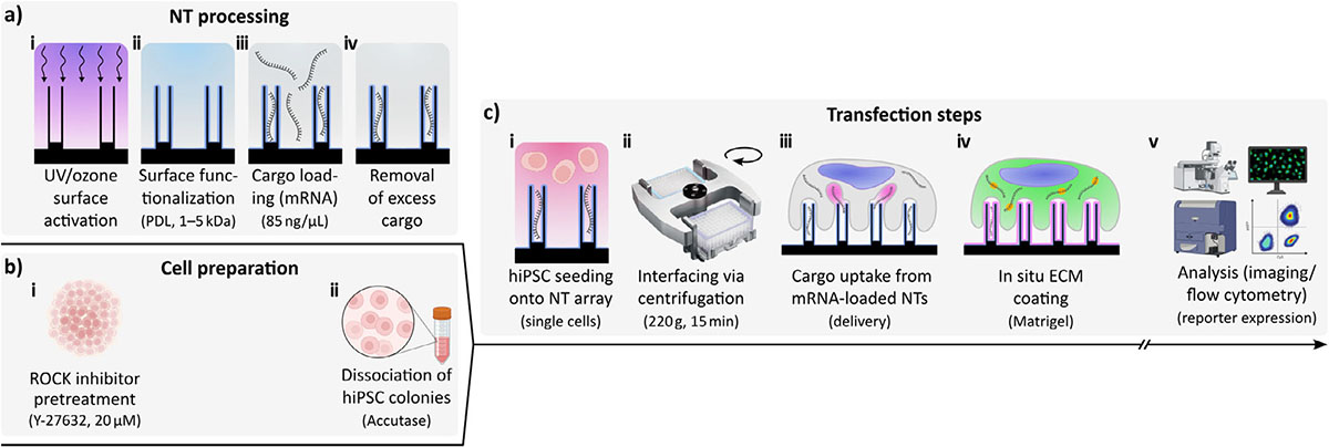 |
| — Workflow of the nanotube-mediated cargo delivery of mRNA to hiPSCs. Briefly, hiPSCs were interfaced with mRNA-loaded NTs and analyzed the next day for the expression of a reporter protein (mCherry, GFP, YPet). a-i) Surface activation with UV/ozone. a-ii) Surface functionalization with poly-D-lysine (PDL, mol. wt.: 1–5 kDa). a-iii) NT loading with mRNA (85 ng/µL). a-iv) Removal of the cargo supernatant. b-i) ROCK inhibitor preconditioning of the hiPSCs with 20 µM Y-27632. b-ii) Dissociation of the hiPSC colonies using Accutase. c-i) hiPSC seeding onto the NT array (single cells). c-ii) Centrifugation of cells onto NTs (220 g, 15 min). c-iii) Cargo uptake from mRNA-loaded NTs. c-iv) In situ extracellular matrix (ECM) coating (Matrigel). c-v) Analysis of expression using confocal microscopy imaging and flow cytometry (reporter proteins). (Image: Reproduced from DOI10.1002/adma.202521046:, CC BY) (click on image to enlarge) |
| The platform uses silicon nanotube arrays fabricated through deep reactive ion etching, a controlled plasma-based process that carves vertical structures from silicon wafers. The nanoscale ring patterns that define each tube were written by electron-beam lithography, which uses a focused beam of electrons to draw features with nanometer precision. The nanotubes are spaced so that each cell sits on roughly 20 of them, and each one tapers to a rim thinner than 50 nm at the tip. |
| That sharp-edge geometry matters. When a cell membrane drapes over a feature with curvature below 200 nm, it triggers endocytosis, the process by which cells actively internalize material from their surroundings. Solid nanoneedles of the same dimensions but without a hollow interior produced no transfection, confirming the importance of both the sharp rim and the internal reservoir. |
| The hollow cavity creates a storage volume 7 to 10 times larger than previous nanotube designs. To attract negatively charged mRNA into these reservoirs, the surfaces were coated with short-chain poly-D-lysine (1 to 5 kDa), a positively charged molecule. Molecular weight proved critical. Standard high-molecular-weight poly-D-lysine (30 to 70 kDa) bound the mRNA too tightly, preventing its release inside cells and collapsing transfection to roughly 3%. |
| Two workflow adaptations made nanoinjection compatible with hiPSC biology. The first involved timing the application of Matrigel, an extracellular matrix gel that hiPSCs need for adhesion and long-term survival. When Matrigel was present from the start, transfection fell below 1%. The matrix proteins likely trapped the mRNA or introduced RNA-degrading enzyme contamination. By seeding cells onto loaded nanotubes first and adding Matrigel one hour later, the team separated cargo uptake from matrix coating. |
| The second adaptation addressed cell spreading. A pretreatment with a ROCK inhibitor, a small molecule that relaxes the cell's internal scaffolding by suppressing a tension-generating signaling pathway, caused hiPSCs to flatten and spread, promoting tighter contact with nanotube tips. Gentle centrifugation immediately after seeding locked the interface in place. Even a five-minute delay in centrifugation cut transfection to roughly 34%. |
| The method delivered three different reporter mRNAs, mCherry, GFP, and YPet, with comparable efficiency and bright, evenly distributed fluorescence throughout the cytoplasm. Co-delivery of mCherry and GFP yielded double-positive cells at roughly 61%, with the two signals tracking each other closely, meaning cells that received more of one mRNA also received proportionally more of the other. Cell viability on the nanotube arrays was approximately 75%, matching flat silicon controls. |
| Nanoinjected hiPSCs maintained their stem cell character after harvesting. They reformed compact colonies and continued to express NANOG, OCT4, and SOX2, three key genes that confirm stem cell identity, at close to 100% across three passages spanning roughly three weeks. When directed toward a neuronal fate using a chemically triggered differentiation protocol, cells from both passage one and passage three produced neurons expressing established neuronal markers at levels matching untreated controls. |
| Because the platform uses mRNA rather than DNA, it avoids genomic integration entirely, sidestepping a persistent concern in both research and clinical translation. RNA is already proving its versatility across vaccines, gene therapy, and diagnostics, and efficient delivery into difficult cell types like hiPSCs expands that reach further. The co-transfection capability opens the door to simultaneously delivering multiple transcription factors or components of gene-editing systems such as CRISPR. |
| With the nanotube arrays already fabricated at wafer level, scaling to the cell numbers required for therapeutic manufacturing is within reach. The work offers a concrete path toward engineering human stem cells without compromising the cellular integrity on which all downstream applications depend. |
 By
Michael
Berger
– Michael is author of four books by the Royal Society of Chemistry:
Nano-Society: Pushing the Boundaries of Technology (2009),
Nanotechnology: The Future is Tiny (2016),
Nanoengineering: The Skills and Tools Making Technology Invisible (2019), and
Waste not! How Nanotechnologies Can Increase Efficiencies Throughout Society (2025)
Copyright ©
Nanowerk LLC
By
Michael
Berger
– Michael is author of four books by the Royal Society of Chemistry:
Nano-Society: Pushing the Boundaries of Technology (2009),
Nanotechnology: The Future is Tiny (2016),
Nanoengineering: The Skills and Tools Making Technology Invisible (2019), and
Waste not! How Nanotechnologies Can Increase Efficiencies Throughout Society (2025)
Copyright ©
Nanowerk LLC
|
|
|
|
ORCID information
|

Become a Spotlight guest author! Join our large and growing group of guest contributors. Have you just published a scientific paper or have other exciting developments to share with the nanotechnology community? Here is how to publish on nanowerk.com. |
